


 Foundational characteristics of cancer include proliferation, angiogenesis, migration, evasion of apoptosis, and cellular immortality. Find key markers for these cellular processes and antibodies to detect them.
 The SUMOplot™ Analysis Program predicts and scores sumoylation sites in your protein. SUMOylation is a post-translational modification involved in various cellular processes, such as nuclear-cytosolic transport, transcriptional regulation, apoptosis, protein stability, response to stress, and progression through the cell cycle.
 The Autophagy Receptor Motif Plotter predicts and scores autophagy receptor binding sites in your protein. Identifying proteins connected to this pathway is critical to understanding the role of autophagy in physiological as well as pathological processes such as development, differentiation, neurodegenerative diseases, stress, infection, and cancer.
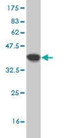
MFGE8 Antibody (monoclonal) (M09)
Mouse monoclonal antibody raised against a partial recombinant MFGE8.
- SPECIFICATION
- CITATIONS
- PROTOCOLS
- BACKGROUND

Application
| WB, E |
|---|---|
| Primary Accession | Q08431 |
| Other Accession | NM_005928 |
| Reactivity | Human |
| Host | Mouse |
| Clonality | Monoclonal |
| Isotype | IgG2a Kappa |
| Clone Names | 3E10 |
| Calculated MW | 43105 Da |
| Gene ID | 4240 |
|---|---|
| Other Names | Lactadherin, Breast epithelial antigen BA46, HMFG, MFGM, Milk fat globule-EGF factor 8, MFG-E8, SED1, Lactadherin short form, Medin, MFGE8 |
| Target/Specificity | MFGE8 (NP_005919, 61 a.a. ~ 170 a.a) partial recombinant protein with GST tag. MW of the GST tag alone is 26 KDa. |
| Dilution | WB~~1:500~1000 E~~N/A |
| Format | Clear, colorless solution in phosphate buffered saline, pH 7.2 . |
| Storage | Store at -20°C or lower. Aliquot to avoid repeated freezing and thawing. |
| Precautions | MFGE8 Antibody (monoclonal) (M09) is for research use only and not for use in diagnostic or therapeutic procedures. |

Thousands of laboratories across the world have published research that depended on the performance of antibodies from Abcepta to advance their research. Check out links to articles that cite our products in major peer-reviewed journals, organized by research category.
info@abcepta.com, and receive a free "I Love Antibodies" mug.

Provided below are standard protocols that you may find useful for product applications.
References
Aberrant splicing of the milk fat globule-EGF factor 8 (MFG-E8) gene in human systemic lupus erythematosus. Yamaguchi H, et al. Eur J Immunol, 2010 Jun. PMID 20213738.Sequential use of transcriptional profiling, expression quantitative trait mapping, and gene association implicates MMP20 in human kidney aging. Wheeler HE, et al. PLoS Genet, 2009 Oct. PMID 19834535.Genetic polymorphism in milk fat globule-EGF factor 8 (MFG-E8) is associated with systemic lupus erythematosus in human. Hu CY, et al. Lupus, 2009 Jul. PMID 19502262.Milk fat globule protein epidermal growth factor-8: a pivotal relay element within the angiotensin II and monocyte chemoattractant protein-1 signaling cascade mediating vascular smooth muscle cells invasion. Fu Z, et al. Circ Res, 2009 Jun 19. PMID 19443842.SED1/MFG-E8: a bi-motif protein that orchestrates diverse cellular interactions. Raymond A, et al. J Cell Biochem, 2009 Apr 15. PMID 19204935.
If you have used an Abcepta product and would like to share how it has performed, please click on the "Submit Review" button and provide the requested information. Our staff will examine and post your review and contact you if needed.
If you have any additional inquiries please email technical services at tech@abcepta.com.
Ordering Information
Other Products
Shipping Information
