


 Foundational characteristics of cancer include proliferation, angiogenesis, migration, evasion of apoptosis, and cellular immortality. Find key markers for these cellular processes and antibodies to detect them.
 The SUMOplot™ Analysis Program predicts and scores sumoylation sites in your protein. SUMOylation is a post-translational modification involved in various cellular processes, such as nuclear-cytosolic transport, transcriptional regulation, apoptosis, protein stability, response to stress, and progression through the cell cycle.
 The Autophagy Receptor Motif Plotter predicts and scores autophagy receptor binding sites in your protein. Identifying proteins connected to this pathway is critical to understanding the role of autophagy in physiological as well as pathological processes such as development, differentiation, neurodegenerative diseases, stress, infection, and cancer.
PCGF2 Antibody (monoclonal) (M04)
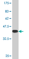
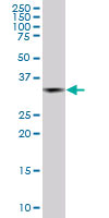
Mouse monoclonal antibody raised against a partial recombinant PCGF2.
- SPECIFICATION
- CITATIONS
- PROTOCOLS
- BACKGROUND

Application
| WB |
|---|---|
| Primary Accession | P35227 |
| Other Accession | BC004858 |
| Reactivity | Human, Rat |
| Host | mouse |
| Clonality | Monoclonal |
| Isotype | IgG2a Kappa |
| Clone Names | 4C10 |
| Calculated MW | 37788 Da |
| Gene ID | 7703 |
|---|---|
| Other Names | Polycomb group RING finger protein 2, DNA-binding protein Mel-18, RING finger protein 110, Zinc finger protein 144, PCGF2, MEL18, RNF110, ZNF144 |
| Target/Specificity | PCGF2 (AAH04858.1, 236 a.a. ~ 294 a.a) partial recombinant protein with GST tag. MW of the GST tag alone is 26 KDa. |
| Dilution | WB~~1:500~1000 |
| Format | Clear, colorless solution in phosphate buffered saline, pH 7.2 . |
| Storage | Store at -20°C or lower. Aliquot to avoid repeated freezing and thawing. |
| Precautions | PCGF2 Antibody (monoclonal) (M04) is for research use only and not for use in diagnostic or therapeutic procedures. |

Thousands of laboratories across the world have published research that depended on the performance of antibodies from Abcepta to advance their research. Check out links to articles that cite our products in major peer-reviewed journals, organized by research category.
info@abcepta.com, and receive a free "I Love Antibodies" mug.

Provided below are standard protocols that you may find useful for product applications.
Background
The protein encoded by this gene contains a RING finger motif and is similar to the polycomb group (PcG) gene products. PcG gene products form complexes via protein-protein interaction and maintain the transcription repression of genes involved in embryogenesis, cell cycles, and tumorigenesis. This protein was shown to act as a negative regulator of transcription and has tumor suppressor activity. The expression of this gene was detected in various tumor cells, but is limited in neural organs in normal tissues. Knockout studies in mice suggested that this protein may negatively regulate the expression of different cytokines, chemokines, and chemokine receptors, and thus plays an important role in lymphocyte differentiation and migration, as well as in immune responses.
References
BMI1 and Mel-18 oppositely regulate carcinogenesis and progression of gastric cancer. Zhang XW, et al. Mol Cancer, 2010 Feb 21. PMID 20170541.The novel tumor-suppressor Mel-18 in prostate cancer: its functional polymorphism, expression and clinical significance. Wang W, et al. Int J Cancer, 2009 Dec 15. PMID 19585577.Mel-18 interacts with RanGAP1 and inhibits its sumoylation. Zhang J, et al. Biochem Biophys Res Commun, 2008 Oct 17. PMID 18706886.A phosphorylated form of Mel-18 targets the Ring1B histone H2A ubiquitin ligase to chromatin. Elderkin S, et al. Mol Cell, 2007 Oct 12. PMID 17936708.Violating the splicing rules: TG dinucleotides function as alternative 3' splice sites in U2-dependent introns. Szafranski K, et al. Genome Biol, 2007. PMID 17672918.
If you have used an Abcepta product and would like to share how it has performed, please click on the "Submit Review" button and provide the requested information. Our staff will examine and post your review and contact you if needed.
If you have any additional inquiries please email technical services at tech@abcepta.com.
Ordering Information
Other Products
Shipping Information
