


 Foundational characteristics of cancer include proliferation, angiogenesis, migration, evasion of apoptosis, and cellular immortality. Find key markers for these cellular processes and antibodies to detect them.
 The SUMOplot™ Analysis Program predicts and scores sumoylation sites in your protein. SUMOylation is a post-translational modification involved in various cellular processes, such as nuclear-cytosolic transport, transcriptional regulation, apoptosis, protein stability, response to stress, and progression through the cell cycle.
 The Autophagy Receptor Motif Plotter predicts and scores autophagy receptor binding sites in your protein. Identifying proteins connected to this pathway is critical to understanding the role of autophagy in physiological as well as pathological processes such as development, differentiation, neurodegenerative diseases, stress, infection, and cancer.
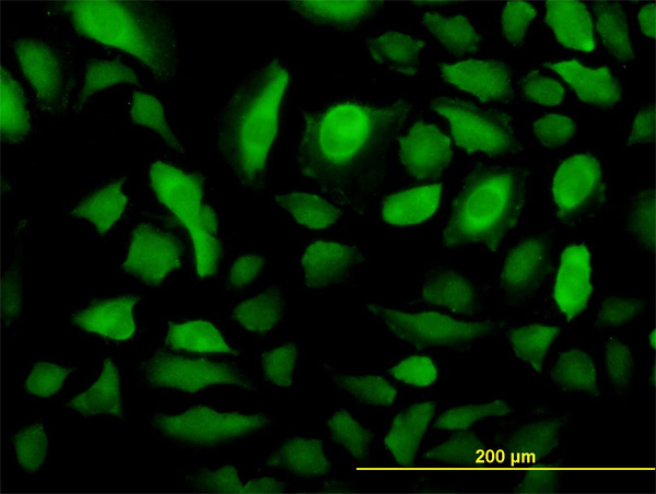
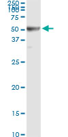
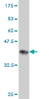
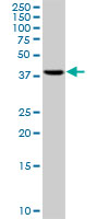
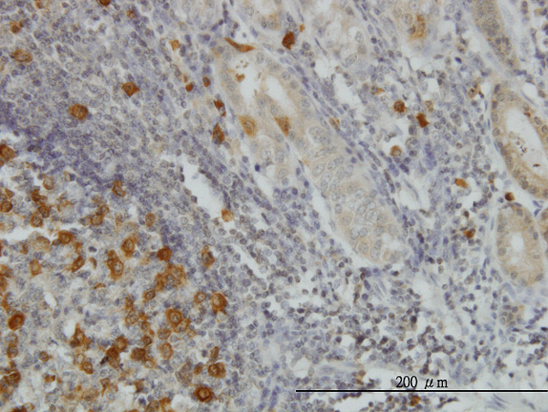
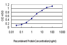
RRM2 Antibody (monoclonal) (M01)
Mouse monoclonal antibody raised against a partial recombinant RRM2.
- SPECIFICATION
- CITATIONS
- PROTOCOLS
- BACKGROUND

Application
| WB, IHC, IF, IP, E |
|---|---|
| Primary Accession | P31350 |
| Other Accession | NM_001034 |
| Reactivity | Human |
| Host | Mouse |
| Clonality | Monoclonal |
| Isotype | IgG1 Kappa |
| Clone Names | 10 |
| Calculated MW | 44878 Da |
| Gene ID | 6241 |
|---|---|
| Other Names | Ribonucleoside-diphosphate reductase subunit M2, Ribonucleotide reductase small chain, Ribonucleotide reductase small subunit, RRM2, RR2 |
| Target/Specificity | RRM2 (NP_001025, 1 a.a. ~ 110 a.a) partial recombinant protein with GST tag. MW of the GST tag alone is 26 KDa. |
| Dilution | WB~~1:500~1000 IHC~~1:100~500 IF~~1:50~200 IP~~N/A E~~N/A |
| Format | Clear, colorless solution in phosphate buffered saline, pH 7.2 . |
| Storage | Store at -20°C or lower. Aliquot to avoid repeated freezing and thawing. |
| Precautions | RRM2 Antibody (monoclonal) (M01) is for research use only and not for use in diagnostic or therapeutic procedures. |

Thousands of laboratories across the world have published research that depended on the performance of antibodies from Abcepta to advance their research. Check out links to articles that cite our products in major peer-reviewed journals, organized by research category.
info@abcepta.com, and receive a free "I Love Antibodies" mug.

Provided below are standard protocols that you may find useful for product applications.
Background
This gene encodes one of two non-identical subunits for ribonucleotide reductase. This reductase catalyzes the formation of deoxyribonucleotides from ribonucleotides. Synthesis of the encoded protein (M2) is regulated in a cell-cycle dependent fashion. Transcription from this gene can initiate from alternative promoters, which results in two isoforms that differ in the lengths of their N-termini. Related pseudogenes have been identified on chromosomes 1 and X.
References
1.RRM1 maintains centrosomal integrity via CHK1 and CDK1 signaling during replication stress.Kim SH, Park ER, Joo HY, Shen YN, Hong SH, Kim CH, Singh R, Lee KH, Shin HJCancer Lett. 2014 Jan 14. pii: S0304-3835(14)00014-7. doi: 10.1016/j.canlet.2013.12.031.2.KRAS-mediated Up-regulation of RRM2 Expression Is Essential for the Proliferation of Colorectal Cancer Cell Lines.Yoshida Y, Tsunoda T, Doi K, Tanaka Y, Fujimoto T, Machida T, Ota T, Koyanagi M, Takashima Y, Sasazuki T, Kuroki M, Iwasaki A, Shirasawa S.Anticancer Research July 2011 vol. 31 no. 7 2535-25393.Epstein-Barr virus episome stability is coupled to a delay in replication timing.Zhou J, Snyder A, Lieberman PM.J Virol. 2009 Mar;83(5):2154-62. Epub 2008 Dec 10.4.Quantitative phosphoproteome profiling of Wnt3a mediated signaling network: indicating the involvement of ribonucleoside-diphosphate reductase M2 subunit phosphorylation at residue serine-20 in canonical Wnt signal transduction.Tang LY, Deng N, Wang LS, Dai J, Wang ZL, Jiang XS, Li SJ, Li L, Sheng QH, Wu DQ, Li L, Zeng R.Mol Cell Proteomics. 2007 Nov;6(11):1952-67. Epub 2007 Aug 12.
If you have used an Abcepta product and would like to share how it has performed, please click on the "Submit Review" button and provide the requested information. Our staff will examine and post your review and contact you if needed.
If you have any additional inquiries please email technical services at tech@abcepta.com.
Ordering Information
Other Products
Shipping Information
