


 Foundational characteristics of cancer include proliferation, angiogenesis, migration, evasion of apoptosis, and cellular immortality. Find key markers for these cellular processes and antibodies to detect them.
 The SUMOplot™ Analysis Program predicts and scores sumoylation sites in your protein. SUMOylation is a post-translational modification involved in various cellular processes, such as nuclear-cytosolic transport, transcriptional regulation, apoptosis, protein stability, response to stress, and progression through the cell cycle.
 The Autophagy Receptor Motif Plotter predicts and scores autophagy receptor binding sites in your protein. Identifying proteins connected to this pathway is critical to understanding the role of autophagy in physiological as well as pathological processes such as development, differentiation, neurodegenerative diseases, stress, infection, and cancer.
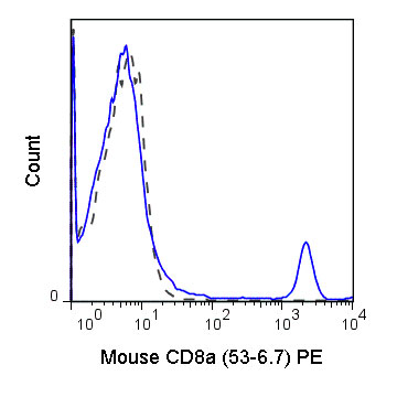
PE Anti-Mouse CD8a (53-6.7) Antibody
- SPECIFICATION
- CITATIONS
- PROTOCOLS
- BACKGROUND

Application
| FC |
|---|---|
| Isotype | Rat IgG2a, kappa |
| Concentration | 0.2 mg/mL |
| Reactivity | Mouse |
| Formulation | 10 mM NaH2PO4, 150 mM NaCl, 0.09% NaN3, 0.1% gelatin, pH7.2 |
| Host | Rat |
| Gene ID | 12525 |
|---|---|
| Gene Name | Cd8a |
| Alternative Name(s) | CD8 alpha, Ly-2, Ly-35, Ly-B, Lyt-2 |
| Format | PE |
| Preparation | This monoclonal antibody was purified from tissue culture supernatant via affinity chromatography. The purified antibody was conjugated under optimal conditions, with unreacted dye removed from the preparation. It is recommended to store the product undiluted at 4°C, and protected from prolonged exposure to light. Do not freeze. |
| Application Notes | This antibody preparation has been quality-tested for flow cytometry using mouse spleen cells, or an appropriate cell type (where indicated). The amount of antibody required for optimal staining of a cell sample should be determined empirically in your system. |
| Storage Conditions | 2-8°C protected from light |
Willinger T and Flavell RA. 2012. Proc. Natl. Acad. Sci. 109:8670-8675. (Flow cytometry)
Thaventhiran JED, Hoffmann A, Magiera L, de la Roche M, Lingel H, Brunner-Weinzierl M, and Fearon DT. 2012. Proc. Natl. Acad. Sci. 10.1073. (Immunohistochemistry – OCT embedded frozen tissue)
Mochimaru H, Usui T, Yaguchi T, Nagahama Y, Hasegawa G, Usui Y, Shimmura S, Tsubota K, Amano S, Kawakami Y, and Ishida S. 2008. Invest. Ophthalmol. Vis. Sci. 49(5):2172-2127. (in vivo cell depletion)
Fan K, Zhou M, Pathak MK, Lindner DJ, Altuntas CZ, Touhy VK, Borden EC, and Yi T. 2005. J. Immunol. 175:7003-7008. (Immunohistochemistry – frozen tissue)
Nutt SL, Metcalf D, D’Amico A, Polli M, and Wu L. 2005. J. Exp. Med. 201:221-231. (Immunomagnetic bead depletion)
Fan G-C, and Singh, RR. 2002. J. Exp. Med. 196: 731-741. (in vitro cell depletion)
Bosselut R, Zhang W, Ashe JM, Kopacz JL, Samelson LE, and Singer A. 1999. J. Exp. Med. 190: 1517-1526. (Immunoprecipitation)
Provided below are standard protocols that you may find useful for product applications.
Background
The 53-6.7 antibody reacts with the 32-34 kDa alpha subunit of mouse CD8, known as CD8a or CD8 alpha. CD8a can form a homodimer (CD8 alpha-alpha), but is more commonly expressed as a heterodimer with a second chain known as CD8b or CD8 beta. CD8 acts as a co-receptor in antigen recognition and subsequent T cell activation that is initiated upon binding of the T cell receptor (TCR) to antigen-bearing MHC Class I molecules. The cytoplasmic domains of CD8 provide binding sites for the tyrosine kinase lck, facilitating intracellular signaling events that lead to T cell activation, development, and cytotoxic effector functions. CD8+ cytotoxic T cells (CTLs) play an important role in inducing cell death of tumor cells, as well as cells infected by virus, bacteria or parasites.
The 53-6.7 antibody is widely used as a phenotypic marker for mouse CD8a expression on cytotoxic T cells, thymocytes, as well as on certain cell types that do not also express the TCR, including some NK cells and lymphoid dendritic cells.

If you have used an Abcepta product and would like to share how it has performed, please click on the "Submit Review" button and provide the requested information. Our staff will examine and post your review and contact you if needed.
If you have any additional inquiries please email technical services at tech@abcepta.com.
Ordering Information
Other Products
Shipping Information
