


 Foundational characteristics of cancer include proliferation, angiogenesis, migration, evasion of apoptosis, and cellular immortality. Find key markers for these cellular processes and antibodies to detect them.
 The SUMOplot™ Analysis Program predicts and scores sumoylation sites in your protein. SUMOylation is a post-translational modification involved in various cellular processes, such as nuclear-cytosolic transport, transcriptional regulation, apoptosis, protein stability, response to stress, and progression through the cell cycle.
 The Autophagy Receptor Motif Plotter predicts and scores autophagy receptor binding sites in your protein. Identifying proteins connected to this pathway is critical to understanding the role of autophagy in physiological as well as pathological processes such as development, differentiation, neurodegenerative diseases, stress, infection, and cancer.
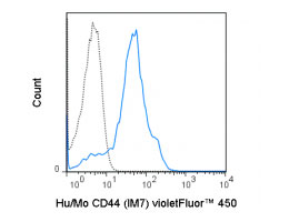
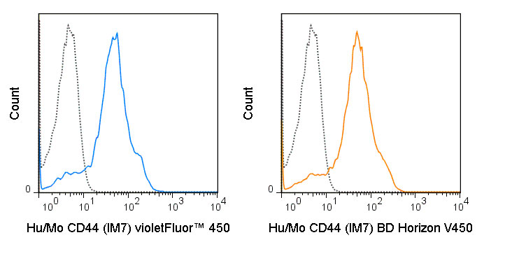
violetFluor™ 450 Anti-Human/Mouse CD44 (IM7) Antibody
- SPECIFICATION
- CITATIONS
- PROTOCOLS
- BACKGROUND

Application
| FC |
|---|---|
| Isotype | Rat IgG2b, kappa |
| Concentration | 0.2 mg/mL |
| Reactivity | Human, Mouse |
| Formulation | 10 mM NaH2PO4, 150 mM NaCl, 0.09% NaN3, 0.1% gelatin, pH7.2 |
| Gene ID | 12505 |
|---|---|
| Gene Name | Cd44 |
| Alternative Name(s) | Pgp-1, MDU3, Hermes, Hyaluronate receptor |
| Format | violetFluor™ 450 |
| Preparation | This monoclonal antibody was purified from tissue culture supernatant via affinity chromatography. The purified antibody was conjugated under optimal conditions, with unreacted dye removed from the preparation. It is recommended to store the product undiluted at 4°C, and protected from prolonged exposure to light. Do not freeze. |
| Application Notes | This antibody preparation has been quality-tested for flow cytometry using mouse spleen cells, or an appropriate cell type (where indicated). The amount of antibody required for optimal staining of a cell sample should be determined empirically in your system. violetFluor™ 450 dye is excited by the violet (405 nm) laser and has a peak emission of 450 nm. The most common band pass filters for this dye are 440/40 or 450/50. violetFluor™ 450 can be used as an alternative for Pacific Blue®, BD Horizon™ V450 or eFluor® 450. |
| Storage Conditions | 2-8°C protected from light |
Chandler HL, Haeussler DJ, Gemensky-Metzler AJ, Wilkie DA, and Lutz EA. 2012. Invest. Ophthalmol. Vis. Sci. 53:1835-1845. (in vitro blocking, canine)
Lee L-F, Logronio K, Tu GH, Zhai W, Ni I, Mei L, Dilley J, Yu J, et al. 2012. Proc. Natl. Acad. Sci. 10.1073. (Flow cytometry).
Ruffell B, Poon GFT, Lee SSM, Brown KL, Tjew S-L, Cooper J, and Johnson P. 2011. J. Biol. Chem. 286:19179-19190. (Immunoprecipitation)
Miyake Y, Matsumoto H, Yokoo M, Miyazawa K et al. 2006. Biol. Reprod. 74: 501-510. (Immunohistochemistry – frozen tissue, swine)
Veir JK, Lappin MR, and Dow SW. 2006. Journal of Feline Medicine and Surgery. 8:400-411. (Flow cytometry – feline)
Frank NY, Margaryan A, Huang Y, Schatton T, Waaga-Gasser AM, Gasser M, Sayegh MH, Sadee W, and Frank MH. 2005. Cancer Res. 65:4320-4333. (Immunohistochemistry – frozen tissue)
Fischer A, Schumacher N, Maier M, Sendtner M, and Gessler M. 2004. Genes & Dev. 18:901-911. (Immunohistochemistry – paraffin embedded tissue)
Xu H, Manivannan A, Liversidge J, Sharp PF, Forrester JV, and Crane IJ. 2002. J. Leukoc. Biol. 72:1133-1141. (in vivo functional assays, induction of apoptosis)
Si-Tahar M, Sitaraman S, Shibahara T, and Madara JL. 2001. Am. J. Physiol. Cell Physiol. 280:C423-C432. (in vitro functional assays, Western Blot
Provided below are standard protocols that you may find useful for product applications.
Background
The IM7 antibody recognizes CD44, a ubiquitously expressed cell surface receptor which is important for extracellular matrix organization, cell-cell and cell-matrix adhesion and migration. CD44 may be expressed in a number of different isoforms (splice variants) from the most typical or “standard” form, known as CD44s, to variants designated CD44v, e.g. CD44v1 or CD44v6. These receptors interact with several ligands, but most often associate with an extracellular matrix component hyaluronate, through which it mediates adhesion.
The IM7 antibody may be used for detection of all isoforms of CD44, as it recognizes constant epitopes near the extracellular proximal domain. (Xu et al, 2002, J. Leukoc. Biol. 72:1133-1141). It has been reported to be cross-reactive with many non-human species including Baboon, Chimpanzee, Cynomolgus, Rhesus, Horse, Cow, Pig, Dog and Cat CD44.

If you have used an Abcepta product and would like to share how it has performed, please click on the "Submit Review" button and provide the requested information. Our staff will examine and post your review and contact you if needed.
If you have any additional inquiries please email technical services at tech@abcepta.com.
Ordering Information
Shipping Information
