


 Foundational characteristics of cancer include proliferation, angiogenesis, migration, evasion of apoptosis, and cellular immortality. Find key markers for these cellular processes and antibodies to detect them.
 The SUMOplot™ Analysis Program predicts and scores sumoylation sites in your protein. SUMOylation is a post-translational modification involved in various cellular processes, such as nuclear-cytosolic transport, transcriptional regulation, apoptosis, protein stability, response to stress, and progression through the cell cycle.
 The Autophagy Receptor Motif Plotter predicts and scores autophagy receptor binding sites in your protein. Identifying proteins connected to this pathway is critical to understanding the role of autophagy in physiological as well as pathological processes such as development, differentiation, neurodegenerative diseases, stress, infection, and cancer.
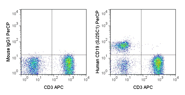
PerCP Anti-Human CD19 (SJ25C1) Antibody
- SPECIFICATION
- CITATIONS
- PROTOCOLS
- BACKGROUND

Application
| FC |
|---|---|
| Isotype | Mouse IgG1, kappa |
| Concentration | 5 µL (0.25 µg)/test |
| Reactivity | Human |
| Formulation | 10 mM NaH2PO4, 150 mM NaCl, 0.09% NaN3, 0.1% gelatin, pH7.2 |
| Gene ID | 930 |
|---|---|
| Gene Name | CD19 |
| Alternative Name(s) | Leu-12, B4 |
| Format | PerCP |
| Storage Conditions | 2-8°C protected from light |
Provided below are standard protocols that you may find useful for product applications.
Background
The SJ25C1 antibody reacts with human CD19, a 95 kDa glycoprotein which acts as a co-receptor, along with CD21 (CR2), CD81 (TAPA-1) and CD225 (Leu13), in support of the functional B cell receptor (BCR). This complex provides antigen-specific recognition and subsequent activation of B cells to proliferate and differentiate into antibody-secreting cells (plasma cells) or memory B cells, which are crucial for secondary antigen encounter. Upon activation and tyrosine phosphorylation, the CD19 molecule can provide an anchor for cytoplasmic signaling proteins such as GRB2, SOS or PLCG2. CD19 is a lineage-differentiation marker, as its expression is detectable at the earliest B cell stages, through development, and is finally lost upon transition to mature plasma cells. The SJ25C1 antibody is widely used as a phenotypic marker for CD19 expression on B cells, as well as on dendritic cell subsets.
References
Piatosa B, Birbach M, Siewiera K, Ussowicz M, Kalwak K, Drabko K, Rekawek A, Tkaczyk K, Kurowski PN. 2013. Cytometry Part B. 84B: 179–186. (Flow Cytometry)
Sekiguchi DR, Smith SB, Sutter JA, Goodman NG, Propert K, Louzoun Y, Rogers W, Luning Prak ET. 2011. Cytometry Part B. 80B: 291–299. (Flow Cytometry)
Dörken B, Möller P, Pezzutto A, Schwartz-Albiez R, Moldenhauer G. B-cell antigens: CD19. In: Knapp W, Dörken B, Gilks WR, et al, ed. Leucocyte Typing IV: White Cell Differentiation Antigens. New York, NY: Oxford University Press; 1989:34-36. (Flow Cytometry)
Karahan GE, Eikmans M, Anholts JDH, Class FHJ and Heidt S. 2014. Clin Exp Immunol. 177(1): 333-340. (Flow Cytometry)
Bansal RR, Mackay CR, Moser B and Ebert M. 2012. Eur J Immunol. 42(1): 110-119. (Flow Cytometry)
Jourdan M, Caraux A, Caron G, Robert N, Fiol G, Rème T, Bolloré K, Vendrell JP, Le Gallou S, Mourcin F, De Vos J, Kassambara A, Duperray C, Hose D, Fest T, Tarte K and Klein B. 2011. J Immunol. 187(8): 3931-3941. (Flow Cytometry)
Goval J-J, Greimers R, Boniver J and de Leval L. 2006. J Histochem Cytochem. 54(1): 75-84. (Immunofluorescence – frozen tissue)

If you have used an Abcepta product and would like to share how it has performed, please click on the "Submit Review" button and provide the requested information. Our staff will examine and post your review and contact you if needed.
If you have any additional inquiries please email technical services at tech@abcepta.com.
Ordering Information
Other Products
Shipping Information
