


 Foundational characteristics of cancer include proliferation, angiogenesis, migration, evasion of apoptosis, and cellular immortality. Find key markers for these cellular processes and antibodies to detect them.
 The SUMOplot™ Analysis Program predicts and scores sumoylation sites in your protein. SUMOylation is a post-translational modification involved in various cellular processes, such as nuclear-cytosolic transport, transcriptional regulation, apoptosis, protein stability, response to stress, and progression through the cell cycle.
 The Autophagy Receptor Motif Plotter predicts and scores autophagy receptor binding sites in your protein. Identifying proteins connected to this pathway is critical to understanding the role of autophagy in physiological as well as pathological processes such as development, differentiation, neurodegenerative diseases, stress, infection, and cancer.
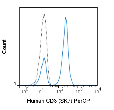
PerCP Anti-Human CD3 (SK7) Antibody
- SPECIFICATION
- CITATIONS
- PROTOCOLS
- BACKGROUND

Application
| FC |
|---|---|
| Isotype | Mouse IgG1, kappa |
| Concentration | 5 µL (0.25 µg)/test |
| Reactivity | Human |
| Formulation | 10 mM NaH2PO4, 150 mM NaCl, 0.09% NaN3, 0.1% gelatin, pH7.2 |
| Gene ID | 916 |
|---|---|
| Gene Name | CD3E |
| Alternative Name(s) | Leu-4, T3 |
| Format | PerCP |
| Storage Conditions | 2-8°C protected from light |
Provided below are standard protocols that you may find useful for product applications.
Background
The SK7 antibody is specific for human CD3e, also known as CD3 epsilon, a 20 kDa subunit of the T cell receptor complex, along with CD3 gamma and CD3 delta. These integral membrane protein chains assemble with additional chains of the T cell receptor (TCR), as well as CD3 zeta chain, to form the T cell receptor – CD3 complex. Together with co-receptors CD4 or CD8, the complex serves to recognize antigens bound to MHC molecules on antigen-presenting cells. These interactions promote T cell receptor signaling (T cell activation), inducing cell proliferation, differentiation, production of cytokines or activation-induced cell death. CD3 is differentially expressed during thymocyte-to-T cell development and on all mature T cells. The SK7 antibody is a widely used phenotypic marker for human T cells. This antibody may induce T cell activation in the presence of monocytes. The antibody has also been demonstrated to be cross-reactive with Chimpanzee CD3. Binding of clone SK7 can be blocked by an alternative Anti-Human CD3 clone, OKT3. Please choose the appropriate format for each application.

If you have used an Abcepta product and would like to share how it has performed, please click on the "Submit Review" button and provide the requested information. Our staff will examine and post your review and contact you if needed.
If you have any additional inquiries please email technical services at tech@abcepta.com.
Ordering Information
Other Products
Shipping Information
