


 Foundational characteristics of cancer include proliferation, angiogenesis, migration, evasion of apoptosis, and cellular immortality. Find key markers for these cellular processes and antibodies to detect them.
 The SUMOplot™ Analysis Program predicts and scores sumoylation sites in your protein. SUMOylation is a post-translational modification involved in various cellular processes, such as nuclear-cytosolic transport, transcriptional regulation, apoptosis, protein stability, response to stress, and progression through the cell cycle.
 The Autophagy Receptor Motif Plotter predicts and scores autophagy receptor binding sites in your protein. Identifying proteins connected to this pathway is critical to understanding the role of autophagy in physiological as well as pathological processes such as development, differentiation, neurodegenerative diseases, stress, infection, and cancer.
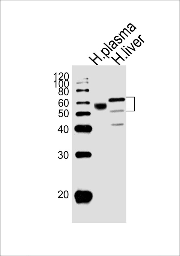
AHSG Antibody (C-term)
Affinity Purified Rabbit Polyclonal Antibody (Pab)
- SPECIFICATION
- CITATIONS: 1
- PROTOCOLS
- BACKGROUND

Application
| WB |
|---|---|
| Primary Accession | P02765 |
| Other Accession | NP_001613.2 |
| Reactivity | Human |
| Host | Rabbit |
| Clonality | Polyclonal |
| Calculated MW | H=39 KDa |
| Isotype | Rabbit IgG |
| Antigen Source | HUMAN |
| Gene ID | 197 |
|---|---|
| Antigen Region | 247-276 aa |
| Other Names | AHSG; FETUA; Alpha-2-HS-glycoprotein; Alpha-2-Z-globulin; Ba-alpha-2-glycoprotein; Fetuin-A; Alpha-2-HS-glycoprotein chain A; Alpha-2-HS-glycoprotein chain B |
| Dilution | WB~~1:1000 |
| Target/Specificity | This AHSG antibody is generated from rabbits immunized with a KLH conjugated synthetic peptide between 247-276 amino acids from the C-terminal region of human AHSG. |
| Format | Purified polyclonal antibody supplied in PBS with 0.09% (W/V) sodium azide. This antibody is purified through a protein A column, followed by peptide affinity purification. |
| Storage | Maintain refrigerated at 2-8°C for up to 2 weeks. For long term storage store at -20°C in small aliquots to prevent freeze-thaw cycles. |
| Precautions | AHSG Antibody (C-term) is for research use only and not for use in diagnostic or therapeutic procedures. |
| Name | AHSG |
|---|---|
| Synonyms | FETUA |
| Function | Promotes endocytosis, possesses opsonic properties and influences the mineral phase of bone. Shows affinity for calcium and barium ions. |
| Cellular Location | Secreted. |
| Tissue Location | Synthesized in liver and selectively concentrated in bone matrix. Secreted in plasma. It is also found in dentin in much higher quantities than other plasma proteins |

Provided below are standard protocols that you may find useful for product applications.
Background
Alpha2-HS glycoprotein (AHSG), a glycoprotein present in the serum, is synthesized by hepatocytes. The AHSG molecule consists of two polypeptide chains, which are both cleaved from a proprotein encoded from a single mRNA. It is involved in several functions, such as endocytosis, brain development and the formation of bone tissue. The protein is commonly present in the cortical plate of the immature cerebral cortex and bone marrow hemopoietic matrix, and it has therefore been postulated that it participates in the development of the tissues. However, its exact significance is still obscure.
References
Bailey, S.D., et al. Diabetes Care 33(10):2250-2253(2010) Verduijn, M., et al. Nephrol. Dial. Transplant. (2010) In press : Wang, Y., et al. Zhonghua Yi Xue Yi Chuan Xue Za Zhi 27(3):310-315(2010) Voigt, M., et al. Histopathology 56(6):775-788(2010) Kusnierz-Cabala, B., et al. Clin. Lab. 56 (5-6), 191-195 (2010) :
If you have used an Abcepta product and would like to share how it has performed, please click on the "Submit Review" button and provide the requested information. Our staff will examine and post your review and contact you if needed.
If you have any additional inquiries please email technical services at tech@abcepta.com.
Ordering Information
Other Products
Shipping Information
