


 Foundational characteristics of cancer include proliferation, angiogenesis, migration, evasion of apoptosis, and cellular immortality. Find key markers for these cellular processes and antibodies to detect them.
 The SUMOplot™ Analysis Program predicts and scores sumoylation sites in your protein. SUMOylation is a post-translational modification involved in various cellular processes, such as nuclear-cytosolic transport, transcriptional regulation, apoptosis, protein stability, response to stress, and progression through the cell cycle.
 The Autophagy Receptor Motif Plotter predicts and scores autophagy receptor binding sites in your protein. Identifying proteins connected to this pathway is critical to understanding the role of autophagy in physiological as well as pathological processes such as development, differentiation, neurodegenerative diseases, stress, infection, and cancer.
MESDC2 Antibody (C-term)
Affinity Purified Rabbit Polyclonal Antibody (Pab)
- SPECIFICATION
- CITATIONS
- PROTOCOLS
- BACKGROUND

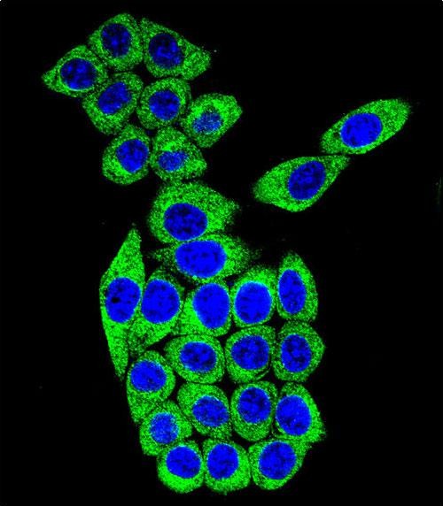
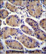
Application
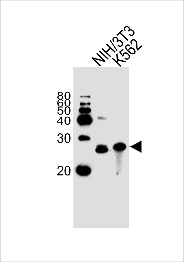
| IF, IHC-P, WB |
|---|---|
| Primary Accession | Q14696 |
| Other Accession | NP_055969.1 |
| Reactivity | Human, Mouse |
| Predicted | Rat |
| Host | Rabbit |
| Clonality | Polyclonal |
| Calculated MW | H=26;M=25;Rat=25 KDa |
| Isotype | Rabbit IgG |
| Antigen Source | HUMAN |
| Gene ID | 23184 |
|---|---|
| Antigen Region | 206-234 aa |
| Other Names | MESDC2; KIAA0081; MESD; LDLR chaperone MESD; Mesoderm development candidate 2; Mesoderm development protein; Renal carcinoma antigen NY-REN-61 |
| Dilution | IF~~1:10~50 IHC-P~~1:10~50 WB~~1:1000 |
| Target/Specificity | This MESDC2 antibody is generated from rabbits immunized with a KLH conjugated synthetic peptide between 206-234 amino acids from the C-terminal region of human MESDC2. |
| Format | Purified polyclonal antibody supplied in PBS with 0.09% (W/V) sodium azide. This antibody is purified through a protein A column, followed by peptide affinity purification. |
| Storage | Maintain refrigerated at 2-8°C for up to 2 weeks. For long term storage store at -20°C in small aliquots to prevent freeze-thaw cycles. |
| Precautions | MESDC2 Antibody (C-term) is for research use only and not for use in diagnostic or therapeutic procedures. |
| Name | MESD (HGNC:13520) |
|---|---|
| Synonyms | KIAA0081, MESDC2, MESDM |
| Function | Chaperone specifically assisting the folding of beta- propeller/EGF modules within the family of low-density lipoprotein receptors (LDLRs) (PubMed:15014448). Acts as a modulator of the Wnt pathway through chaperoning the coreceptors of the canonical Wnt pathway, LRP5 and LRP6, to the plasma membrane (PubMed:17488095, PubMed:23572575). Essential for specification of embryonic polarity and mesoderm induction. Plays an essential role in neuromuscular junction (NMJ) formation by promoting cell-surface expression of LRP4 (By similarity). May regulate phagocytosis of apoptotic retinal pigment epithelium (RPE) cells (By similarity). |
| Cellular Location | Endoplasmic reticulum Note=Released from apoptotic cells and shed photoreceptor outer segments. {ECO:0000250|UniProtKB:Q9ERE7} |

Thousands of laboratories across the world have published research that depended on the performance of antibodies from Abcepta to advance their research. Check out links to articles that cite our products in major peer-reviewed journals, organized by research category.
info@abcepta.com, and receive a free "I Love Antibodies" mug.

Provided below are standard protocols that you may find useful for product applications.
Background
Chaperone specifically assisting the folding of beta-propeller/EGF modules within the family of low-density lipoprotein receptors (LDLRs). Acts as a modulator of the Wnt pathway through chaperoning the coreceptors of the canonical Wnt pathway, LRP5 and LRP6, to the plasma membrane. Essential for specification of embryonic polarity and mesoderm induction.
References
Murrills, R.J., et al. J. Cell. Biochem. 108(5):1066-1075(2009)
Li, Y., et al. FEBS Lett. 580(22):5423-5428(2006)
Veltman, I.M., et al. Hum. Mol. Genet. 14(14):1955-1963(2005)
Clark, H.F., et al. Genome Res. 13(10):2265-2270(2003)
Hsieh, J.C., et al. Cell 112(3):355-367(2003)
If you have used an Abcepta product and would like to share how it has performed, please click on the "Submit Review" button and provide the requested information. Our staff will examine and post your review and contact you if needed.
If you have any additional inquiries please email technical services at tech@abcepta.com.
Ordering Information
Shipping Information
