


 Foundational characteristics of cancer include proliferation, angiogenesis, migration, evasion of apoptosis, and cellular immortality. Find key markers for these cellular processes and antibodies to detect them.
 The SUMOplot™ Analysis Program predicts and scores sumoylation sites in your protein. SUMOylation is a post-translational modification involved in various cellular processes, such as nuclear-cytosolic transport, transcriptional regulation, apoptosis, protein stability, response to stress, and progression through the cell cycle.
 The Autophagy Receptor Motif Plotter predicts and scores autophagy receptor binding sites in your protein. Identifying proteins connected to this pathway is critical to understanding the role of autophagy in physiological as well as pathological processes such as development, differentiation, neurodegenerative diseases, stress, infection, and cancer.
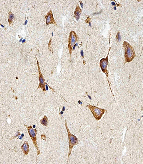
PLAUR Antibody (C-term)
Purified Rabbit Polyclonal Antibody (Pab)
- SPECIFICATION
- CITATIONS
- PROTOCOLS
- BACKGROUND

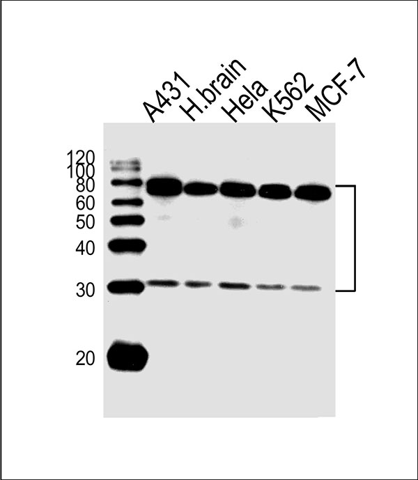
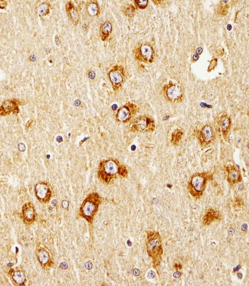
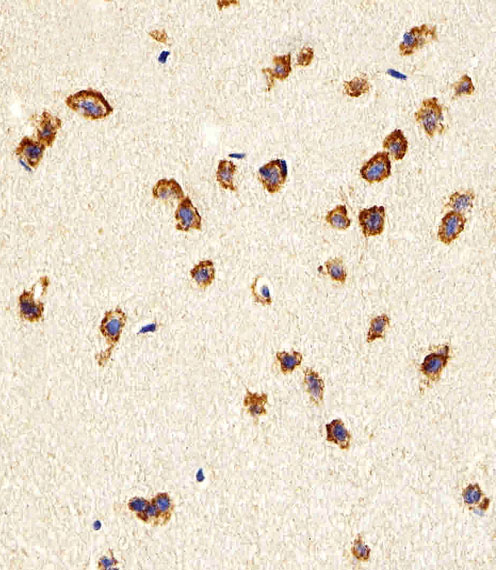
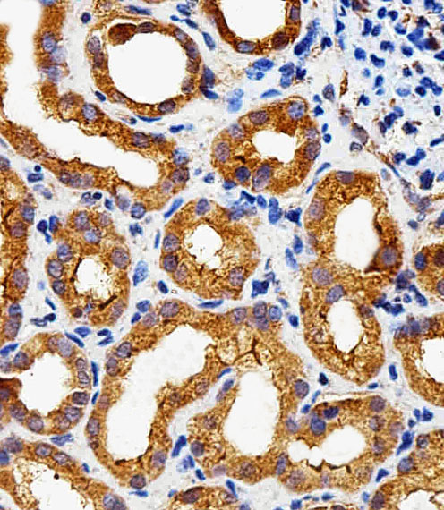
Application
| WB, IHC-P |
|---|---|
| Primary Accession | Q03405 |
| Reactivity | Human |
| Host | Rabbit |
| Clonality | Polyclonal |
| Calculated MW | H=37,32 KDa |
| Isotype | Rabbit IgG |
| Antigen Source | HUMAN |
| Gene ID | 5329 |
|---|---|
| Antigen Region | 273-305 aa |
| Other Names | Urokinase plasminogen activator surface receptor, U-PAR, uPAR, Monocyte activation antigen Mo3, CD87, PLAUR, MO3, UPAR |
| Dilution | WB~~1:1000 IHC-P~~1:25 |
| Target/Specificity | This PLAUR antibody is generated from a rabbit immunized with a KLH conjugated synthetic peptide between 273-305 amino acids from the C-terminal region of human PLAUR. |
| Format | Purified polyclonal antibody supplied in PBS with 0.09% (W/V) sodium azide. This antibody is purified through a protein A column, followed by peptide affinity purification. |
| Storage | Maintain refrigerated at 2-8°C for up to 2 weeks. For long term storage store at -20°C in small aliquots to prevent freeze-thaw cycles. |
| Precautions | PLAUR Antibody (C-term) is for research use only and not for use in diagnostic or therapeutic procedures. |
| Name | PLAUR |
|---|---|
| Synonyms | MO3, UPAR |
| Function | Acts as a receptor for urokinase plasminogen activator (PubMed:15677461). Plays a role in localizing and promoting plasmin formation. Mediates the proteolysis-independent signal transduction activation effects of U-PA. It is subject to negative-feedback regulation by U-PA which cleaves it into an inactive form. |
| Cellular Location | Cell membrane. Cell projection, invadopodium membrane Note=Colocalized with FAP (seprase) preferentially at the cell surface of invadopodia membrane in a cytoskeleton-, integrin- and vitronectin- dependent manner. [Isoform 2]: Secreted {ECO:0000250|UniProtKB:P49616} |
| Tissue Location | Expressed in neurons of the rolandic area of the brain (at protein level). Expressed in the brain |

Thousands of laboratories across the world have published research that depended on the performance of antibodies from Abcepta to advance their research. Check out links to articles that cite our products in major peer-reviewed journals, organized by research category.
info@abcepta.com, and receive a free "I Love Antibodies" mug.

Provided below are standard protocols that you may find useful for product applications.
Background
Acts as a receptor for urokinase plasminogen activator. Plays a role in localizing and promoting plasmin formation. Mediates the proteolysis-independent signal transduction activation effects of U-PA. It is subject to negative-feedback regulation by U-PA which cleaves it into an inactive form.
References
Roldan A.L.,et al.EMBO J. 9:467-474(1990).
Min H.Y.,et al.J. Immunol. 148:3636-3642(1992).
Bayraktutan U.,et al.Biochem. Soc. Trans. 21:395-395(1993).
Pyke C.,et al.FEBS Lett. 326:69-74(1993).
Casey J.R.,et al.Blood 84:1151-1156(1994).
If you have used an Abcepta product and would like to share how it has performed, please click on the "Submit Review" button and provide the requested information. Our staff will examine and post your review and contact you if needed.
If you have any additional inquiries please email technical services at tech@abcepta.com.
Ordering Information
Other Products
Shipping Information
