


 Foundational characteristics of cancer include proliferation, angiogenesis, migration, evasion of apoptosis, and cellular immortality. Find key markers for these cellular processes and antibodies to detect them.
 The SUMOplot™ Analysis Program predicts and scores sumoylation sites in your protein. SUMOylation is a post-translational modification involved in various cellular processes, such as nuclear-cytosolic transport, transcriptional regulation, apoptosis, protein stability, response to stress, and progression through the cell cycle.
 The Autophagy Receptor Motif Plotter predicts and scores autophagy receptor binding sites in your protein. Identifying proteins connected to this pathway is critical to understanding the role of autophagy in physiological as well as pathological processes such as development, differentiation, neurodegenerative diseases, stress, infection, and cancer.
HLA-DRB1 Antibody (Center)
Purified Rabbit Polyclonal Antibody (Pab)
- SPECIFICATION
- CITATIONS
- PROTOCOLS
- BACKGROUND

Application
| WB |
|---|---|
| Primary Accession | P01911 |
| Other Accession | P13762, Q29974, Q30167 |
| Reactivity | Human |
| Host | Rabbit |
| Clonality | polyclonal |
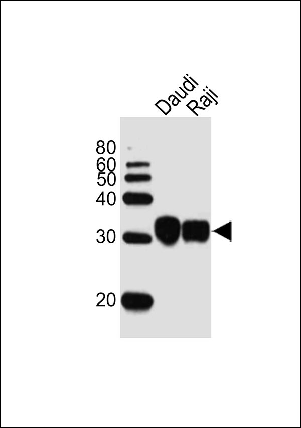
| Calculated MW | H=30 KDa |
| Isotype | Rabbit IgG |
| Antigen Source | HUMAN |
| Gene ID | 3123 |
|---|---|
| Antigen Region | 122-154 aa |
| Other Names | HLA class II histocompatibility antigen, DRB1-15 beta chain, DW22/DR22, MHC class II antigen DRB1*15, HLA-DRB1, HLA-DRB2 |
| Dilution | WB~~1:1000 |
| Target/Specificity | This HLA-DRB1 antibody is generated from a rabbit immunized with a KLH conjugated synthetic peptide between 122-154 amino acids from the Central region of human HLA-DRB1. |
| Format | Purified polyclonal antibody supplied in PBS with 0.09% (W/V) sodium azide. This antibody is purified through a protein A column, followed by peptide affinity purification. |
| Storage | Maintain refrigerated at 2-8°C for up to 2 weeks. For long term storage store at -20°C in small aliquots to prevent freeze-thaw cycles. |
| Precautions | HLA-DRB1 Antibody (Center) is for research use only and not for use in diagnostic or therapeutic procedures. |
| Name | HLA-DRB1 (HGNC:4948) |
|---|---|
| Function | A beta chain of antigen-presenting major histocompatibility complex class II (MHCII) molecule. In complex with the alpha chain HLA- DRA, displays antigenic peptides on professional antigen presenting cells (APCs) for recognition by alpha-beta T cell receptor (TCR) on HLA-DRB1-restricted CD4-positive T cells. This guides antigen-specific T-helper effector functions, both antibody-mediated immune response and macrophage activation, to ultimately eliminate the infectious agents and transformed cells (PubMed:15265931, PubMed:16148104, PubMed:22327072, PubMed:27591323, PubMed:29884618, PubMed:31495665, PubMed:8642306). Typically presents extracellular peptide antigens of 10 to 30 amino acids that arise from proteolysis of endocytosed antigens in lysosomes (PubMed:8145819). In the tumor microenvironment, presents antigenic peptides that are primarily generated in tumor- resident APCs likely via phagocytosis of apoptotic tumor cells or macropinocytosis of secreted tumor proteins (PubMed:31495665). Presents peptides derived from intracellular proteins that are trapped in autolysosomes after macroautophagy, a mechanism especially relevant for T cell selection in the thymus and central immune tolerance (PubMed:17182262, PubMed:23783831). The selection of the immunodominant epitopes follows two processing modes: 'bind first, cut/trim later' for pathogen-derived antigenic peptides and 'cut first, bind later' for autoantigens/self-peptides (PubMed:25413013). The anchor residue at position 1 of the peptide N-terminus, usually a large hydrophobic residue, is essential for high affinity interaction with MHCII molecules (PubMed:8145819). |
| Cellular Location | Cell membrane; Single-pass type I membrane protein. Endoplasmic reticulum membrane; Single-pass type I membrane protein. Lysosome membrane; Single-pass type I membrane protein. Late endosome membrane; Single-pass type I membrane protein. Autolysosome membrane Note=The MHC class II complex transits through a number of intracellular compartments in the endocytic pathway until it reaches the cell membrane for antigen presentation (PubMed:18305173). Component of immunological synapses at the interface between T cell and APC (PubMed:29884618). |
| Tissue Location | Expressed in professional APCs: monocyte/macrophages, dendritic cells and B cells (at protein level) (PubMed:19830726, PubMed:23783831, PubMed:31495665). Expressed in thymic epithelial cells (at protein level) (PubMed:23783831) |

Thousands of laboratories across the world have published research that depended on the performance of antibodies from Abcepta to advance their research. Check out links to articles that cite our products in major peer-reviewed journals, organized by research category.
info@abcepta.com, and receive a free "I Love Antibodies" mug.

Provided below are standard protocols that you may find useful for product applications.
Background
Binds peptides derived from antigens that access the endocytic route of antigen presenting cells (APC) and presents them on the cell surface for recognition by the CD4 T-cells. The peptide binding cleft accommodates peptides of 10-30 residues. The peptides presented by MHC class II molecules are generated mostly by degradation of proteins that access the endocytic route, where they are processed by lysosomal proteases and other hydrolases. Exogenous antigens that have been endocytosed by the APC are thus readily available for presentation via MHC II molecules, and for this reason this antigen presentation pathway is usually referred to as exogenous. As membrane proteins on their way to degradation in lysosomes as part of their normal turn-over are also contained in the endosomal/lysosomal compartments, exogenous antigens must compete with those derived from endogenous components. Autophagy is also a source of endogenous peptides, autophagosomes constitutively fuse with MHC class II loading compartments. In addition to APCs, other cells of the gastrointestinal tract, such as epithelial cells, express MHC class II molecules and CD74 and act as APCs, which is an unusual trait of the GI tract. To produce a MHC class II molecule that presents an antigen, three MHC class II molecules (heterodimers of an alpha and a beta chain) associate with a CD74 trimer in the ER to form a heterononamer. Soon after the entry of this complex into the endosomal/lysosomal system where antigen processing occurs, CD74 undergoes a sequential degradation by various proteases, including CTSS and CTSL, leaving a small fragment termed CLIP (class-II-associated invariant chain peptide). The removal of CLIP is facilitated by HLA-DM via direct binding to the alpha-beta-CLIP complex so that CLIP is released. HLA-DM stabilizes MHC class II molecules until primary high affinity antigenic peptides are bound. The MHC II molecule bound to a peptide is then transported to the cell membrane surface. In B-cells, the interaction between HLA-DM and MHC class II molecules is regulated by HLA-DO. Primary dendritic cells (DCs) also to express HLA-DO. Lysosomal microenvironment has been implicated in the regulation of antigen loading into MHC II molecules, increased acidification produces increased proteolysis and efficient peptide loading.
References
Lock C.B.,et al.Immunogenetics 27:449-455(1988).
Raymond C.K.,et al.Genome Res. 15:1250-1257(2005).
Balas A.,et al.Hum. Immunol. 67:1008-1016(2006).
Mungall A.J.,et al.Nature 425:805-811(2003).
Wu S.K.,et al.J. Immunol. 138:2953-2959(1987).
If you have used an Abcepta product and would like to share how it has performed, please click on the "Submit Review" button and provide the requested information. Our staff will examine and post your review and contact you if needed.
If you have any additional inquiries please email technical services at tech@abcepta.com.
Ordering Information
Other Products
Shipping Information
