


 Foundational characteristics of cancer include proliferation, angiogenesis, migration, evasion of apoptosis, and cellular immortality. Find key markers for these cellular processes and antibodies to detect them.
 The SUMOplot™ Analysis Program predicts and scores sumoylation sites in your protein. SUMOylation is a post-translational modification involved in various cellular processes, such as nuclear-cytosolic transport, transcriptional regulation, apoptosis, protein stability, response to stress, and progression through the cell cycle.
 The Autophagy Receptor Motif Plotter predicts and scores autophagy receptor binding sites in your protein. Identifying proteins connected to this pathway is critical to understanding the role of autophagy in physiological as well as pathological processes such as development, differentiation, neurodegenerative diseases, stress, infection, and cancer.
FN3KRP Antibody (N-Term)
Purified Rabbit Polyclonal Antibody (Pab)
- SPECIFICATION
- CITATIONS
- PROTOCOLS
- BACKGROUND

Application
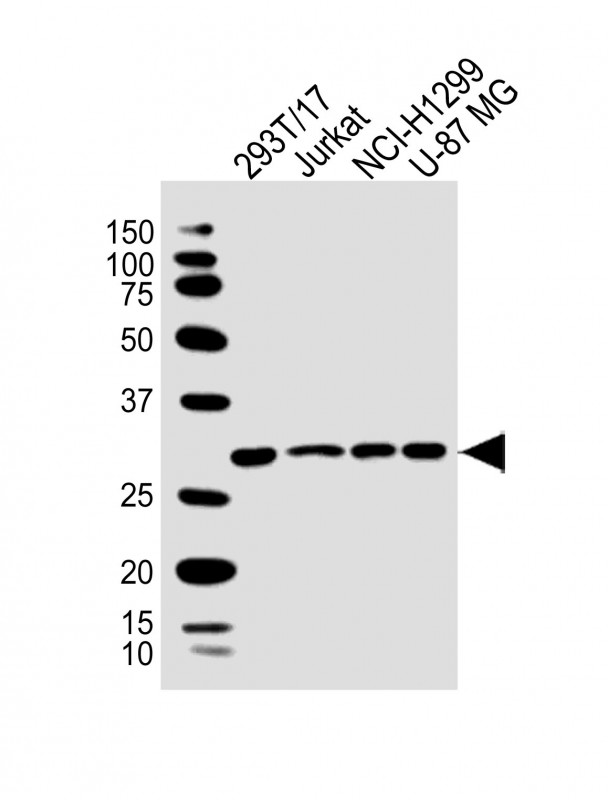
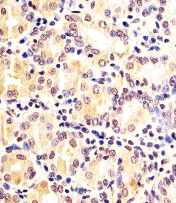
| WB, IHC |
|---|---|
| Primary Accession | Q9HA64 |
| Reactivity | Human |
| Host | Rabbit |
| Clonality | Polyclonal |
| Calculated MW | H=34;M=34 KDa |
| Isotype | Rabbit IgG |
| Antigen Source | HUMAN |
| Gene ID | 79672 |
|---|---|
| Antigen Region | 24-58 aa |
| Other Names | Ketosamine-3-kinase, 271-, Fructosamine-3-kinase-related protein, FN3K-RP, FN3K-related protein, FN3KRP |
| Dilution | WB~~1:2000 IHC~~1:25 |
| Target/Specificity | This FN3KRP antibody is generated from a rabbit immunized with a KLH conjugated synthetic peptide between 24-58 amino acids from human FN3KRP. |
| Storage | Maintain refrigerated at 2-8°C for up to 2 weeks. For long term storage store at -20°C in small aliquots to prevent freeze-thaw cycles. |
| Precautions | FN3KRP Antibody (N-Term) is for research use only and not for use in diagnostic or therapeutic procedures. |
| Name | FN3KRP {ECO:0000303|PubMed:15137908, ECO:0000312|HGNC:HGNC:25700} |
|---|---|
| Function | Ketosamine-3-kinase involved in protein deglycation by mediating phosphorylation of ribuloselysine and psicoselysine on glycated proteins, to generate ribuloselysine-3 phosphate and psicoselysine-3 phosphate, respectively (PubMed:14633848, PubMed:15137908). Ribuloselysine-3 phosphate and psicoselysine-3 phosphate adducts are unstable and decompose under physiological conditions (PubMed:14633848, PubMed:15137908). Not able to phosphorylate fructoselysine (PubMed:14633848). |
| Tissue Location | Widely expressed; except in skeletal muscle where it is expressed at very low level (PubMed:15331600). Expressed in erythrocytes (PubMed:15137908). |

Thousands of laboratories across the world have published research that depended on the performance of antibodies from Abcepta to advance their research. Check out links to articles that cite our products in major peer-reviewed journals, organized by research category.
info@abcepta.com, and receive a free "I Love Antibodies" mug.

Provided below are standard protocols that you may find useful for product applications.
Background
Phosphorylates psicosamines and ribulosamines, but not fructosamines, on the third carbon of the sugar moiety. Protein- bound psicosamine 3-phosphates and ribulosamine 3-phosphates are unstable and decompose under physiological conditions. Thus phosphorylation leads to deglycation.
References
Collard F.,et al.Diabetes 52:2888-2895(2003).
Wiemann S.,et al.Genome Res. 11:422-435(2001).
Ota T.,et al.Nat. Genet. 36:40-45(2004).
Collard F.,et al.Biochem. J. 382:137-143(2004).
Oppermann F.S.,et al.Mol. Cell. Proteomics 8:1751-1764(2009).
If you have used an Abcepta product and would like to share how it has performed, please click on the "Submit Review" button and provide the requested information. Our staff will examine and post your review and contact you if needed.
If you have any additional inquiries please email technical services at tech@abcepta.com.
Ordering Information
Shipping Information
