


 Foundational characteristics of cancer include proliferation, angiogenesis, migration, evasion of apoptosis, and cellular immortality. Find key markers for these cellular processes and antibodies to detect them.
 The SUMOplot™ Analysis Program predicts and scores sumoylation sites in your protein. SUMOylation is a post-translational modification involved in various cellular processes, such as nuclear-cytosolic transport, transcriptional regulation, apoptosis, protein stability, response to stress, and progression through the cell cycle.
 The Autophagy Receptor Motif Plotter predicts and scores autophagy receptor binding sites in your protein. Identifying proteins connected to this pathway is critical to understanding the role of autophagy in physiological as well as pathological processes such as development, differentiation, neurodegenerative diseases, stress, infection, and cancer.
BAP31 Antibody
Purified Mouse Monoclonal Antibody (Mab)
- SPECIFICATION
- CITATIONS
- PROTOCOLS
- BACKGROUND

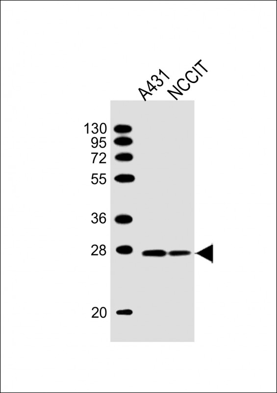
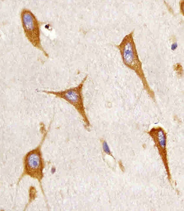
Application
| WB, IHC |
|---|---|
| Primary Accession | P51572 |
| Reactivity | Human |
| Host | Mouse |
| Clonality | Monoclonal |
| Calculated MW | H=28,35;M=28 KDa |
| Isotype | IgG1,Κ |
| Antigen Source | HUMAN |
| Gene ID | 10134 |
|---|---|
| Antigen Region | 122-246 aa |
| Other Names | B-cell receptor-associated protein 31, BCR-associated protein 31, Bap31, 6C6-AG tumor-associated antigen, Protein CDM, p28, BCAP31, BAP31, DXS1357E |
| Dilution | WB~~1:2000 IHC~~1:25 |
| Target/Specificity | This BAP31 antibody is generated from a mouse immunized with a recombinant. |
| Storage | Maintain refrigerated at 2-8°C for up to 2 weeks. For long term storage store at -20°C in small aliquots to prevent freeze-thaw cycles. |
| Precautions | BAP31 Antibody is for research use only and not for use in diagnostic or therapeutic procedures. |
| Name | BCAP31 (HGNC:16695) |
|---|---|
| Function | Functions as a chaperone protein (PubMed:18287538, PubMed:9396746). Is one of the most abundant endoplasmic reticulum (ER) proteins (PubMed:18287538, PubMed:9396746). Plays a role in the export of secreted proteins in the ER, the recognition of abnormally folded protein and their targeting to the ER associated-degradation (ERAD) (PubMed:18287538, PubMed:9396746). Also serves as a cargo receptor for the export of transmembrane proteins (By similarity). Plays a role in the assembly of the mitochondrial membrane respiratory chain NADH dehydrogenase (Complex I) by stimulating the translocation of NDUFS4 and NDUFB11 from the cytosol to the mitochondria via interaction with TOMM40 (PubMed:31206022). In response to ER stress, delocalizes from the ER-mitochondria contact sites and binds BCL2 (PubMed:31206022). May be involved in CASP8-mediated apoptosis (PubMed:10958671). |
| Cellular Location | Endoplasmic reticulum membrane; Multi-pass membrane protein Endoplasmic reticulum-Golgi intermediate compartment membrane; Multi-pass membrane protein. Note=May shuttle between the ER and the intermediate compartment/cis-Golgi complex (PubMed:9396746). Associates with the mitochondria-associated endoplasmic reticulum membrane via interaction with TOMM40 (PubMed:31206022) |
| Tissue Location | Ubiquitous. Highly expressed in neurons and discrete endocrine cells. |

Thousands of laboratories across the world have published research that depended on the performance of antibodies from Abcepta to advance their research. Check out links to articles that cite our products in major peer-reviewed journals, organized by research category.
info@abcepta.com, and receive a free "I Love Antibodies" mug.

Provided below are standard protocols that you may find useful for product applications.
Background
Functions as a chaperone protein. Is one of the most abundant endoplasmic reticulum (ER) proteins. Plays a role in the export of secreted proteins in the ER, the recognition of abnormally folded protein and their targeting to the ER associated-degradation (ERAD). Also serves as a cargo receptor for the export of transmembrane proteins. May be involved in CASP8- mediated apoptosis.
References
Mosser J.,et al.Genomics 22:469-471(1994).
Li E.,et al.Eur. J. Biochem. 238:631-638(1996).
Adachi T.,et al.EMBO J. 15:1534-1541(1996).
Ota T.,et al.Nat. Genet. 36:40-45(2004).
Ross M.T.,et al.Nature 434:325-337(2005).
If you have used an Abcepta product and would like to share how it has performed, please click on the "Submit Review" button and provide the requested information. Our staff will examine and post your review and contact you if needed.
If you have any additional inquiries please email technical services at tech@abcepta.com.
Ordering Information
Other Products
Shipping Information
