


 Foundational characteristics of cancer include proliferation, angiogenesis, migration, evasion of apoptosis, and cellular immortality. Find key markers for these cellular processes and antibodies to detect them.
 The SUMOplot™ Analysis Program predicts and scores sumoylation sites in your protein. SUMOylation is a post-translational modification involved in various cellular processes, such as nuclear-cytosolic transport, transcriptional regulation, apoptosis, protein stability, response to stress, and progression through the cell cycle.
 The Autophagy Receptor Motif Plotter predicts and scores autophagy receptor binding sites in your protein. Identifying proteins connected to this pathway is critical to understanding the role of autophagy in physiological as well as pathological processes such as development, differentiation, neurodegenerative diseases, stress, infection, and cancer.
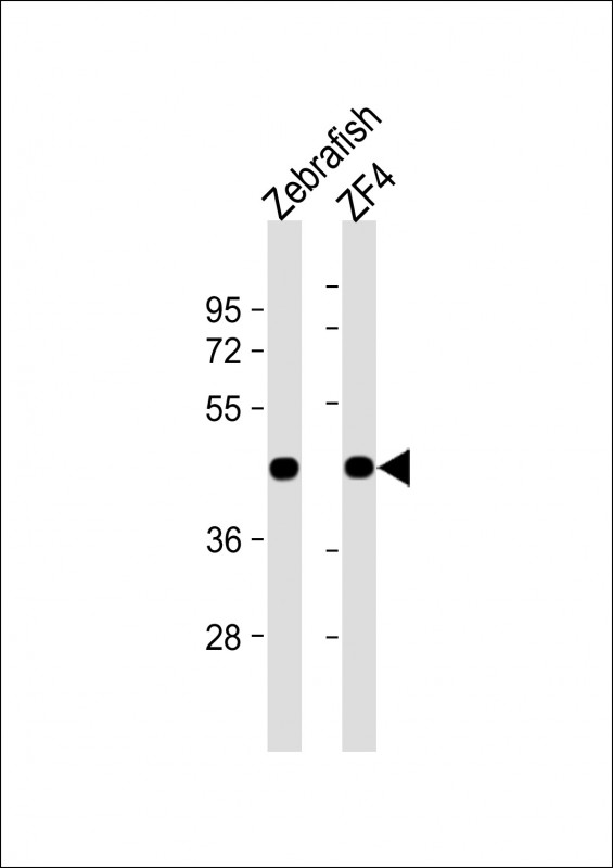
Zebrafish actba Antibody (Center)
Affinity Purified Rabbit Polyclonal Antibody (Pab)
- SPECIFICATION
- CITATIONS
- PROTOCOLS
- BACKGROUND

Application
| WB, E |
|---|---|
| Primary Accession | Q7ZVI7 |
| Other Accession | P60010, P68136, P68135, P68137, P68134, P68133, P68139, P68138, P63269, P63268, P63267, P63270, Q5E9B5, A2BDB0, P63259, P63260, P63261, Q5ZMQ2, P63258, P04751, P68035, P68033, P68032, P68034, Q3ZC07, O93400, P60711, P29751, Q6QAQ1, P60710, Q4R561, P60709 |
| Reactivity | Zebrafish |
| Predicted | C.Elegans, Drosophila, Xenopus, Chicken, Bovine, Human, Mouse, Rabbit, Rat, Hamster, Horse, Monkey, Pig, Sheep, Yeast |
| Host | Rabbit |
| Clonality | Polyclonal |
| Isotype | Rabbit IgG |
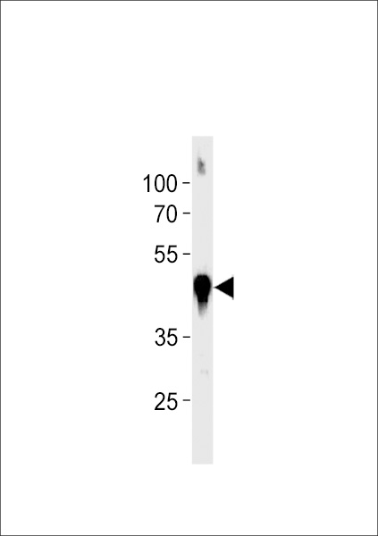
| Calculated MW | 41767 Da |
| Antigen Region | 188-215 aa |
| Gene ID | 57934 |
|---|---|
| Other Names | Actin, cytoplasmic 1, Beta-actin-1, Actin, cytoplasmic 1, N-terminally processed, actba, bact, bactin1, bactzf |
| Target/Specificity | This Zebrafish actba antibody is generated from rabbits immunized with a KLH conjugated synthetic peptide between 188-215 amino acids from the central region of zebrafish actba. |
| Dilution | WB~~1:1000 E~~Use at an assay dependent concentration. |
| Format | Purified polyclonal antibody supplied in PBS with 0.09% (W/V) sodium azide. This antibody is purified through a protein A column, followed by peptide affinity purification. |
| Storage | Maintain refrigerated at 2-8°C for up to 2 weeks. For long term storage store at -20°C in small aliquots to prevent freeze-thaw cycles. |
| Precautions | Zebrafish actba Antibody (Center) is for research use only and not for use in diagnostic or therapeutic procedures. |
| Name | actba |
|---|---|
| Synonyms | bact, bactin1, bactzf |
| Function | Actin is a highly conserved protein that polymerizes to produce filaments that form cross-linked networks in the cytoplasm of cells. Actin exists in both monomeric (G-actin) and polymeric (F-actin) forms, both forms playing key functions, such as cell motility and contraction. In addition to their role in the cytoplasmic cytoskeleton, G- and F-actin also localize in the nucleus, and regulate gene transcription and motility and repair of damaged DNA. |
| Cellular Location | Cytoplasm, cytoskeleton {ECO:0000250|UniProtKB:P60710}. Nucleus {ECO:0000250|UniProtKB:O93400} |
| Tissue Location | Skeletal muscle, heart, gill, digestive tissue and brain (PubMed:9987040). Widespread expression throughout the brain, with highest levels in regions where neuronal proliferation is greatest (PubMed:9987040). |

Thousands of laboratories across the world have published research that depended on the performance of antibodies from Abcepta to advance their research. Check out links to articles that cite our products in major peer-reviewed journals, organized by research category.
info@abcepta.com, and receive a free "I Love Antibodies" mug.

Provided below are standard protocols that you may find useful for product applications.
Background
Actins are highly conserved proteins that are involved in various types of cell motility and are ubiquitously expressed in all eukaryotic cells.
References
Barrallo A., et al. Eur. J. Neurosci. 11:369-372(1999).
If you have used an Abcepta product and would like to share how it has performed, please click on the "Submit Review" button and provide the requested information. Our staff will examine and post your review and contact you if needed.
If you have any additional inquiries please email technical services at tech@abcepta.com.
Ordering Information
Shipping Information
