


 Foundational characteristics of cancer include proliferation, angiogenesis, migration, evasion of apoptosis, and cellular immortality. Find key markers for these cellular processes and antibodies to detect them.
 The SUMOplot™ Analysis Program predicts and scores sumoylation sites in your protein. SUMOylation is a post-translational modification involved in various cellular processes, such as nuclear-cytosolic transport, transcriptional regulation, apoptosis, protein stability, response to stress, and progression through the cell cycle.
 The Autophagy Receptor Motif Plotter predicts and scores autophagy receptor binding sites in your protein. Identifying proteins connected to this pathway is critical to understanding the role of autophagy in physiological as well as pathological processes such as development, differentiation, neurodegenerative diseases, stress, infection, and cancer.
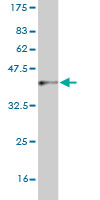
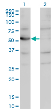
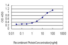
CNDP2 Antibody (monoclonal) (M09)
Mouse monoclonal antibody raised against a partial recombinant CNDP2.
- SPECIFICATION
- CITATIONS: 1
- PROTOCOLS
- BACKGROUND

Application
| WB, E |
|---|---|
| Primary Accession | Q96KP4 |
| Other Accession | NM_018235 |
| Reactivity | Human |
| Host | Mouse |
| Clonality | Monoclonal |
| Isotype | IgG2a Kappa |
| Clone Names | 1B1 |
| Calculated MW | 52878 Da |
| Gene ID | 55748 |
|---|---|
| Other Names | Cytosolic non-specific dipeptidase, CNDP dipeptidase 2, Glutamate carboxypeptidase-like protein 1, Peptidase A, CNDP2, CN2, CPGL, PEPA |
| Target/Specificity | CNDP2 (NP_060705, 191 a.a. ~ 300 a.a) partial recombinant protein with GST tag. MW of the GST tag alone is 26 KDa. |
| Dilution | WB~~1:500~1000 E~~N/A |
| Format | Clear, colorless solution in phosphate buffered saline, pH 7.2 . |
| Storage | Store at -20°C or lower. Aliquot to avoid repeated freezing and thawing. |
| Precautions | CNDP2 Antibody (monoclonal) (M09) is for research use only and not for use in diagnostic or therapeutic procedures. |

Provided below are standard protocols that you may find useful for product applications.
Background
CNDP2, also known as tissue carnosinase and peptidase A (EC 3.4.13.18), is a nonspecific dipeptidase rather than a selective carnosinase (Teufel et al., 2003 [PubMed 12473676]).
References
1.Laser Microdissection and Two-Dimensional Difference Gel Electrophoresis Reveal the Role of a Novel Macrophage-Capping Protein in Lymph Node Metastasis in Gastric Cancer.Ichikawa H, Kanda T, Kosugi SI, Kawachi Y, Sasaki H, Wakai T, Kondo TJ Proteome Res. 2013 Jul 9.2.Proteomic profiling of the substantia nigra demonstrates CNDP2 overexpression in Parkinson's disease.Licker V, Cote M, Lobrinus JA, Rodrigo N, Kovari E, Hochstrasser DF, Turck N, Sanchez JC, Burkhard PR.J Proteomics. 2012 Mar 6. [Epub ahead of print]
If you have used an Abcepta product and would like to share how it has performed, please click on the "Submit Review" button and provide the requested information. Our staff will examine and post your review and contact you if needed.
If you have any additional inquiries please email technical services at tech@abcepta.com.
Ordering Information
Other Products
Shipping Information
