


 Foundational characteristics of cancer include proliferation, angiogenesis, migration, evasion of apoptosis, and cellular immortality. Find key markers for these cellular processes and antibodies to detect them.
 The SUMOplot™ Analysis Program predicts and scores sumoylation sites in your protein. SUMOylation is a post-translational modification involved in various cellular processes, such as nuclear-cytosolic transport, transcriptional regulation, apoptosis, protein stability, response to stress, and progression through the cell cycle.
 The Autophagy Receptor Motif Plotter predicts and scores autophagy receptor binding sites in your protein. Identifying proteins connected to this pathway is critical to understanding the role of autophagy in physiological as well as pathological processes such as development, differentiation, neurodegenerative diseases, stress, infection, and cancer.
CNDP2 Antibody (C-term)
Affinity Purified Rabbit Polyclonal Antibody (Pab)
- SPECIFICATION
- CITATIONS
- PROTOCOLS
- BACKGROUND

Application
| WB, E |
|---|---|
| Primary Accession | Q96KP4 |
| Other Accession | NP_060705.2, NP_001161971.1 |
| Reactivity | Human |
| Host | Rabbit |
| Clonality | Polyclonal |
| Isotype | Rabbit IgG |
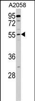
| Calculated MW | 52878 Da |
| Antigen Region | 348-375 aa |
| Gene ID | 55748 |
|---|---|
| Other Names | Cytosolic non-specific dipeptidase, CNDP dipeptidase 2, Glutamate carboxypeptidase-like protein 1, Peptidase A, CNDP2, CN2, CPGL, PEPA |
| Target/Specificity | This CNDP2 antibody is generated from rabbits immunized with a KLH conjugated synthetic peptide between 348-375 amino acids from the C-terminal region of human CNDP2. |
| Dilution | WB~~1:1000 E~~Use at an assay dependent concentration. |
| Format | Purified polyclonal antibody supplied in PBS with 0.09% (W/V) sodium azide. This antibody is purified through a protein A column, followed by peptide affinity purification. |
| Storage | Maintain refrigerated at 2-8°C for up to 2 weeks. For long term storage store at -20°C in small aliquots to prevent freeze-thaw cycles. |
| Precautions | CNDP2 Antibody (C-term) is for research use only and not for use in diagnostic or therapeutic procedures. |
| Name | CNDP2 {ECO:0000303|PubMed:25964343, ECO:0000312|HGNC:HGNC:24437} |
|---|---|
| Function | Catalyzes the peptide bond hydrolysis in dipeptides, displaying a non-redundant activity toward threonyl dipeptides (By similarity). Mediates threonyl dipeptide catabolism in a tissue- specific way (By similarity). Has high dipeptidase activity toward cysteinylglycine, an intermediate metabolite in glutathione metabolism (PubMed:12473676, PubMed:19346245). Metabolizes N-lactoyl-amino acids, both through hydrolysis to form lactic acid and amino acids, as well as through their formation by reverse proteolysis (PubMed:25964343). Plays a role in the regulation of cell cycle arrest and apoptosis (PubMed:17121880, PubMed:24395568). |
| Cellular Location | Cytoplasm |
| Tissue Location | [Isoform 1]: Ubiquitously expressed with higher levels in kidney and liver (at protein level). Expressed in peripheral blood leukocytes (PubMed:12473676). Expressed in gastric mucosa and down-regulated in gastric cancer mucosal tissues (at protein level) (PubMed:24395568). |

Thousands of laboratories across the world have published research that depended on the performance of antibodies from Abcepta to advance their research. Check out links to articles that cite our products in major peer-reviewed journals, organized by research category.
info@abcepta.com, and receive a free "I Love Antibodies" mug.

Provided below are standard protocols that you may find useful for product applications.
Background
CNDP2, also known as tissue carnosinase and peptidase A (EC 3.4.13.18), is a nonspecific dipeptidase rather than a selective carnosinase (Teufel et al., 2003 [PubMed 12473676]).
References
Ahmed, A.H., et al. Biochemistry 49(13):2843-2850(2010)
McDonough, C.W., et al. Hum. Genet. 126(2):265-275(2009)
Wanic, K., et al. Diabetes 57(9):2547-2551(2008)
Wanic, K., et al. Diabetes (2008) In press :
Tu, L.C., et al. Mol. Cell Proteomics 6(4):575-588(2007)
If you have used an Abcepta product and would like to share how it has performed, please click on the "Submit Review" button and provide the requested information. Our staff will examine and post your review and contact you if needed.
If you have any additional inquiries please email technical services at tech@abcepta.com.
Ordering Information
Other Products
Shipping Information
