


 Foundational characteristics of cancer include proliferation, angiogenesis, migration, evasion of apoptosis, and cellular immortality. Find key markers for these cellular processes and antibodies to detect them.
 The SUMOplot™ Analysis Program predicts and scores sumoylation sites in your protein. SUMOylation is a post-translational modification involved in various cellular processes, such as nuclear-cytosolic transport, transcriptional regulation, apoptosis, protein stability, response to stress, and progression through the cell cycle.
 The Autophagy Receptor Motif Plotter predicts and scores autophagy receptor binding sites in your protein. Identifying proteins connected to this pathway is critical to understanding the role of autophagy in physiological as well as pathological processes such as development, differentiation, neurodegenerative diseases, stress, infection, and cancer.
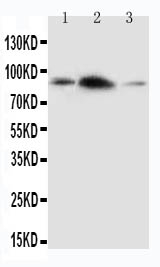
Anti-APLP2 Antibody
- SPECIFICATION
- CITATIONS
- PROTOCOLS
- BACKGROUND

Application
| WB |
|---|---|
| Primary Accession | Q06481 |
| Host | Rabbit |
| Reactivity | Human, Mouse, Rat |
| Clonality | Polyclonal |
| Format | Lyophilized |
| Description | Rabbit IgG polyclonal antibody for Amyloid-like protein 2(APLP2) detection. Tested with WB in Human;Mouse;Rat. |
| Reconstitution | Add 0.2ml of distilled water will yield a concentration of 500ug/ml. |
| Gene ID | 334 |
|---|---|
| Other Names | Amyloid-like protein 2, APLP-2, APPH, Amyloid protein homolog, CDEI box-binding protein, CDEBP, APLP2, APPL2 |
| Calculated MW | 86956 MW KDa |
| Application Details | Western blot, 0.1-0.5 µg/ml, Human, Rat, Mouse |
| Subcellular Localization | Cell membrane ; Single-pass type I membrane protein . Nucleus . |
| Tissue Specificity | Expressed in placenta, brain, heart, lung, liver, kidney and endothelial tissues. |
| Protein Name | Amyloid-like protein 2(APLP-2) |
| Contents | Each vial contains 5mg BSA, 0.9mg NaCl, 0.2mg Na2HPO4, 0.05mg Thimerosal, 0.05mg NaN3. |
| Immunogen | A synthetic peptide corresponding to a sequence at the C-terminus of human APLP2(639-656aa EEKVINSKNKVDENMVID), different from the related rat and mouse sequences by one amino acid. |
| Purification | Immunogen affinity purified. |
| Cross Reactivity | No cross reactivity with other proteins |
| Storage | At -20˚C for one year. After r˚Constitution, at 4˚C for one month. It˚Can also be aliquotted and stored frozen at -20˚C for a longer time.Avoid repeated freezing and thawing. |
| Sequence Similarities | Belongs to the APP family. |
| Name | APLP2 (HGNC:598) |
|---|---|
| Function | May play a role in the regulation of hemostasis. The soluble form may have inhibitory properties towards coagulation factors. May interact with cellular G-protein signaling pathways. May bind to the DNA 5'-GTCACATG-3'(CDEI box). Inhibits trypsin, chymotrypsin, plasmin, factor XIA and plasma and glandular kallikrein. Modulates the Cu/Zn nitric oxide-catalyzed autodegradation of GPC1 heparan sulfate side chains in fibroblasts (By similarity). |
| Cellular Location | Cell membrane; Single-pass type I membrane protein. Nucleus |
| Tissue Location | Expressed in placenta, brain, heart, lung, liver, kidney and endothelial tissues |

Thousands of laboratories across the world have published research that depended on the performance of antibodies from Abcepta to advance their research. Check out links to articles that cite our products in major peer-reviewed journals, organized by research category.
info@abcepta.com, and receive a free "I Love Antibodies" mug.

Provided below are standard protocols that you may find useful for product applications.
Background
APLP2(Amyloid beta(A4) precursor-like protein 2), also known as CDEBP or SPERM MEMBRANE PROTEIN, is a protein that in humans is encoded by the APLP2 gene. The APLP2 gene is mapped on 11q24.3. The human amyloid precursor-like protein APLP2 is a highly conserved homolog of a sequence-specific DNA-binding mouse protein with an important function in the cell cycle. APLP2 along with APLP1 are important modulators of glucose and insulin homeostasis. APLP2 associates with antigen presentation molecules like MHC class I molecules and regulates their surface expression by enhancing endocytosis.
If you have used an Abcepta product and would like to share how it has performed, please click on the "Submit Review" button and provide the requested information. Our staff will examine and post your review and contact you if needed.
If you have any additional inquiries please email technical services at tech@abcepta.com.
Ordering Information
Other Products
Shipping Information
