


 Foundational characteristics of cancer include proliferation, angiogenesis, migration, evasion of apoptosis, and cellular immortality. Find key markers for these cellular processes and antibodies to detect them.
 The SUMOplot™ Analysis Program predicts and scores sumoylation sites in your protein. SUMOylation is a post-translational modification involved in various cellular processes, such as nuclear-cytosolic transport, transcriptional regulation, apoptosis, protein stability, response to stress, and progression through the cell cycle.
 The Autophagy Receptor Motif Plotter predicts and scores autophagy receptor binding sites in your protein. Identifying proteins connected to this pathway is critical to understanding the role of autophagy in physiological as well as pathological processes such as development, differentiation, neurodegenerative diseases, stress, infection, and cancer.
Anti-Bcl2A1 Antibody
- SPECIFICATION
- CITATIONS
- PROTOCOLS
- BACKGROUND

Application
| WB |
|---|---|
| Primary Accession | Q16548 |
| Host | Rabbit |
| Reactivity | Human |
| Clonality | Polyclonal |
| Format | Lyophilized |
| Description | Rabbit IgG polyclonal antibody for Bcl-2-related protein A1(BCL2A1) detection. Tested with WB in Human. |
| Reconstitution | Add 0.2ml of distilled water will yield a concentration of 500ug/ml. |
| Gene ID | 597 |
|---|---|
| Other Names | Bcl-2-related protein A1, Bcl-2-like protein 5, Bcl2-L-5, Hemopoietic-specific early response protein, Protein BFL-1, Protein GRS, BCL2A1, BCL2L5, BFL1, GRS, HBPA1 |
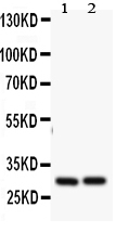
| Calculated MW | 20132 MW KDa |
| Application Details | Western blot, 0.1-0.5 µg/ml, Human |
| Subcellular Localization | Cytoplasm. |
| Tissue Specificity | Seems to be restricted to the hematopoietic compartment. Expressed in peripheral blood, spleen, and bone marrow, at moderate levels in lung, small intestine and testis, at a minimal levels in other tissues. Also found in vascular smooth muscle cells and hematopoietic malignancies. |
| Protein Name | Bcl-2-related protein A1 |
| Contents | Each vial contains 5mg BSA, 0.9mg NaCl, 0.2mg Na2HPO4, 0.05mg Thimerosal, 0.05mg NaN3. |
| Immunogen | A synthetic peptide corresponding to a sequence at the N-terminus of human Bcl2A1(1-16aa MTDCEFGYIYRLAQDY). |
| Purification | Immunogen affinity purified. |
| Cross Reactivity | No cross reactivity with other proteins |
| Storage | At -20˚C for one year. After r˚Constitution, at 4˚C for one month. It˚Can also be aliquotted and stored frozen at -20˚C for a longer time.Avoid repeated freezing and thawing. |
| Name | BCL2A1 |
|---|---|
| Synonyms | BCL2L5, BFL1, GRS, HBPA1 |
| Function | Retards apoptosis induced by IL-3 deprivation. May function in the response of hemopoietic cells to external signals and in maintaining endothelial survival during infection (By similarity). Can inhibit apoptosis induced by serum starvation in the mammary epithelial cell line HC11 (By similarity). |
| Cellular Location | Cytoplasm. |
| Tissue Location | Seems to be restricted to the hematopoietic compartment. Expressed in peripheral blood, spleen, and bone marrow, at moderate levels in lung, small intestine and testis, at a minimal levels in other tissues. Also found in vascular smooth muscle cells and hematopoietic malignancies |

Thousands of laboratories across the world have published research that depended on the performance of antibodies from Abcepta to advance their research. Check out links to articles that cite our products in major peer-reviewed journals, organized by research category.
info@abcepta.com, and receive a free "I Love Antibodies" mug.

Provided below are standard protocols that you may find useful for product applications.
Background
Bcl-2-related protein A1(BCL2A1), is the human homolog of the murine A1, and maps to chromosome 15q24.3. The BCL2A1 gene has also been referred to as BCL2L5, BFL1, and GRS. Bfl-1 protein suppresses apoptosis induced by the p53 tumor suppressor protein in a manner similar to other Bcl-2 family members such as Bcl-2, Bcl-xL and EBV-BHRF1. In addition, the bfl-I gene cooperates efficiently with the Ela oncogene in transformation of primary rodent epithelial cells. The human bfl-1 gene may play an important role in carcinogenesis.
If you have used an Abcepta product and would like to share how it has performed, please click on the "Submit Review" button and provide the requested information. Our staff will examine and post your review and contact you if needed.
If you have any additional inquiries please email technical services at tech@abcepta.com.
Ordering Information
Other Products
Shipping Information
