


 Foundational characteristics of cancer include proliferation, angiogenesis, migration, evasion of apoptosis, and cellular immortality. Find key markers for these cellular processes and antibodies to detect them.
 The SUMOplot™ Analysis Program predicts and scores sumoylation sites in your protein. SUMOylation is a post-translational modification involved in various cellular processes, such as nuclear-cytosolic transport, transcriptional regulation, apoptosis, protein stability, response to stress, and progression through the cell cycle.
 The Autophagy Receptor Motif Plotter predicts and scores autophagy receptor binding sites in your protein. Identifying proteins connected to this pathway is critical to understanding the role of autophagy in physiological as well as pathological processes such as development, differentiation, neurodegenerative diseases, stress, infection, and cancer.
Anti-TXNL2 Picoband Antibody
- SPECIFICATION
- CITATIONS
- PROTOCOLS
- BACKGROUND

Application
| WB |
|---|---|
| Primary Accession | O76003 |
| Host | Rabbit |
| Reactivity | Human, Mouse, Rat |
| Clonality | Polyclonal |
| Format | Lyophilized |
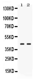
| Description | Rabbit IgG polyclonal antibody for Glutaredoxin-3(GLRX3) detection. Tested with WB in Human;Mouse;Rat. |
| Reconstitution | Add 0.2ml of distilled water will yield a concentration of 500ug/ml. |
| Gene ID | 10539 |
|---|---|
| Other Names | Glutaredoxin-3, PKC-interacting cousin of thioredoxin, PICOT, PKC-theta-interacting protein, PKCq-interacting protein, Thioredoxin-like protein 2, GLRX3, PICOT, TXNL2 |
| Calculated MW | 37432 MW KDa |
| Application Details | Western blot, 0.1-0.5 µg/ml, Mouse, Rat, Human |
| Subcellular Localization | Cytoplasm, cell cortex . Cytoplasm, myofibril, sarcomere, Z line . Under the plasma membrane. After PMA stimulation, GLRX3 and PRKCQ/PKC-theta translocate to a more extended submembrane area. In the Z line, found associated with CSRP3 (By similarity). . |
| Tissue Specificity | Expressed in heart, spleen, testis and, to a lower extent, in thymus and peripheral blood leukocytes. Weakly expressed in lung, placenta, colon and small intestine. |
| Protein Name | Glutaredoxin-3 |
| Contents | Each vial contains 5mg BSA, 0.9mg NaCl, 0.2mg Na2HPO4, 0.05mg NaN3. |
| Immunogen | E. coli-derived human TXNL2 recombinant protein (Position: L89-S177). Human TXNL2 shares 93.3% and 94.4% amino acid (aa) sequence identity with mouse and rat TXNL2, respectively. |
| Purification | Immunogen affinity purified. |
| Cross Reactivity | No cross reactivity with other proteins. |
| Storage | At -20˚C for one year. After r˚Constitution, at 4˚C for one month. It˚Can also be aliquotted and stored frozen at -20˚C for a longer time.Avoid repeated freezing and thawing. |
| Name | GLRX3 |
|---|---|
| Synonyms | PICOT {ECO:0000303|PubMed:10636891}, TXN |
| Function | Together with BOLA2, acts as a cytosolic iron-sulfur (Fe-S) cluster assembly factor that facilitates [2Fe-2S] cluster insertion into a subset of cytosolic proteins (PubMed:26613676, PubMed:27519415). Acts as a critical negative regulator of cardiac hypertrophy and a positive inotropic regulator (By similarity). Required for hemoglobin maturation (PubMed:23615448). Does not possess any thyoredoxin activity since it lacks the conserved motif that is essential for catalytic activity. |
| Cellular Location | Cytoplasm, cytosol. Cytoplasm, cell cortex. Cytoplasm, myofibril, sarcomere, Z line {ECO:0000250|UniProtKB:Q9CQM9}. Note=Under the plasma membrane (By similarity). After PMA stimulation, GLRX3 and PRKCQ/PKC-theta translocate to a more extended submembrane area (By similarity). In the Z line, found associated with CSRP3 (By similarity). {ECO:0000250|UniProtKB:Q9CQM9} |
| Tissue Location | Expressed in heart, spleen, testis and, to a lower extent, in thymus and peripheral blood leukocytes. Weakly expressed in lung, placenta, colon and small intestine |

Thousands of laboratories across the world have published research that depended on the performance of antibodies from Abcepta to advance their research. Check out links to articles that cite our products in major peer-reviewed journals, organized by research category.
info@abcepta.com, and receive a free "I Love Antibodies" mug.

Provided below are standard protocols that you may find useful for product applications.
Background
Glutaredoxin-3 is a protein that in humans is encoded by the GLRX3 gene. This gene encodes a member of the glutaredoxin family. Glutaredoxins are oxidoreductase enzymes that reduce a variety of substrates using glutathione as a cofactor. The encoded protein binds to and modulates the function of protein kinase C theta. The encoded protein may also inhibit apoptosis and play a role in cellular growth, and the expression of this gene may be a marker for cancer. Pseudogenes of this gene are located on the short arm of chromosomes 6 and 9. Alternatively spliced transcript variants have been observed for this gene.
If you have used an Abcepta product and would like to share how it has performed, please click on the "Submit Review" button and provide the requested information. Our staff will examine and post your review and contact you if needed.
If you have any additional inquiries please email technical services at tech@abcepta.com.
Ordering Information
Other Products
Shipping Information
