


 Foundational characteristics of cancer include proliferation, angiogenesis, migration, evasion of apoptosis, and cellular immortality. Find key markers for these cellular processes and antibodies to detect them.
 The SUMOplot™ Analysis Program predicts and scores sumoylation sites in your protein. SUMOylation is a post-translational modification involved in various cellular processes, such as nuclear-cytosolic transport, transcriptional regulation, apoptosis, protein stability, response to stress, and progression through the cell cycle.
 The Autophagy Receptor Motif Plotter predicts and scores autophagy receptor binding sites in your protein. Identifying proteins connected to this pathway is critical to understanding the role of autophagy in physiological as well as pathological processes such as development, differentiation, neurodegenerative diseases, stress, infection, and cancer.
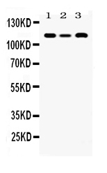
Anti-HLTF Picoband Antibody
- SPECIFICATION
- CITATIONS
- PROTOCOLS
- BACKGROUND

Application
| WB |
|---|---|
| Primary Accession | Q14527 |
| Host | Rabbit |
| Reactivity | Human, Mouse, Rat |
| Clonality | Polyclonal |
| Format | Lyophilized |
| Description | Rabbit IgG polyclonal antibody for Helicase-like transcription factor(HLTF) detection. Tested with WB in Human;Mouse;Rat. |
| Reconstitution | Add 0.2ml of distilled water will yield a concentration of 500ug/ml. |
| Gene ID | 6596 |
|---|---|
| Other Names | Helicase-like transcription factor, 2.3.2.27, 3.6.4.-, DNA-binding protein/plasminogen activator inhibitor 1 regulator, HIP116, RING finger protein 80, RING-type E3 ubiquitin transferase HLTF, SWI/SNF-related matrix-associated actin-dependent regulator of chromatin subfamily A member 3, Sucrose nonfermenting protein 2-like 3, HLTF, HIP116A, RNF80, SMARCA3, SNF2L3, ZBU1 |
| Calculated MW | 113929 MW KDa |
| Application Details | Western blot, 0.1-0.5 µg/ml, Human, Mouse, Rat |
| Subcellular Localization | Cytoplasm . Nucleus . Nucleus, nucleolus . Nucleus, nucleoplasm . Nuclear localization is stimulated by progesterone. . |
| Tissue Specificity | Expressed in brain, heart, kidney, liver, lung, pancreas, placenta and skeletal muscle. . |
| Protein Name | Helicase-like transcription factor |
| Contents | Each vial contains 5mg BSA, 0.9mg NaCl, 0.2mg Na2HPO4, 0.05mg NaN3. |
| Immunogen | E. coli-derived human HLTF recombinant protein (Position: S911-L1009). Human HLTF shares 92.9% amino acid (aa) sequence identity with mouse HLTF. |
| Purification | Immunogen affinity purified. |
| Cross Reactivity | No cross reactivity with other proteins. |
| Storage | At -20˚C for one year. After r˚Constitution, at 4˚C for one month. It˚Can also be aliquotted and stored frozen at -20˚C for a longer time.Avoid repeated freezing and thawing. |
| Name | HLTF (HGNC:11099) |
|---|---|
| Function | Functions as a DNA-dependent ATPase and E3 ubiquitin-protein ligase involved in chromatin regulation and DNA damage tolerance (DDT) (PubMed:18316726, PubMed:18719106, PubMed:26051180, PubMed:31960921, PubMed:39142279, PubMed:40680746). Catalyzes 'Lys-63'-linked polyubiquitination of monoubiquitinated PCNA at 'Lys-164' in response to genotoxic stress, promoting error-free postreplication repair via template switching (PubMed:18316726, PubMed:18719106). Acts as an epigenetic regulator by promoting recruitment of DNMT1, thereby ensuring DNA methylation inheritance: specifically binds histone H3 trimethylated at 'Lys-9' (H3K9me3) and mediates histone H3 'Lys-23' polyubiquitination (H3K23ub), a docking site for DNMT1, leading to DNMT1 recruitment and replication-coupled DNA methylation maintenance (PubMed:40680746). Catalyzes formation of H3K23ub in two steps: first mediates monoubiquitination together with UBE2E1 and UBE2D2, and then extends ubiquitin chains via 'Lys-63'-linked ubiquitination together with UBE2N and UBE2V2 (PubMed:40680746). Also acts as a chromatin redodeling factor, thereby regulating transcription (PubMed:10391891, PubMed:1994885, PubMed:9126292). Exhibits ATP-dependent double-stranded DNA (dsDNA) translocase activity but lacks classical helicase activity; mediates replication fork reversal by concertedly unwinding and annealing nascent and parental strands, thereby suppressing DNA synthesis and maintaining genomic stability (PubMed:1994885). Resolves G-quadruplex (G4) DNA structures in cooperation with MSH2, limiting replication stress and G4 accumulation across the cell cycle (PubMed:39142279). Contributes to nucleotide excision repair by evicting lesion-containing oligonucleotides using its HIRAN and ATPase domains (PubMed:26051180). Can displace single-stranded DNA from triplex structures through ATP-dependent dsDNA translocation (PubMed:26051180, PubMed:31960921). Also has protein clearing activity at the stalled replication fork, facilitating restart of DNA replication (PubMed:21795603). |
| Cellular Location | Nucleus. Chromosome |
| Tissue Location | Expressed in brain, heart, kidney, liver, lung, pancreas, placenta and skeletal muscle. |

Thousands of laboratories across the world have published research that depended on the performance of antibodies from Abcepta to advance their research. Check out links to articles that cite our products in major peer-reviewed journals, organized by research category.
info@abcepta.com, and receive a free "I Love Antibodies" mug.

Provided below are standard protocols that you may find useful for product applications.
Background
Helicase-like transcription factor is an enzyme that in humans is encoded by the HLTF gene. This gene encodes a member of the SWI/SNF family. Members of this family have helicase and ATPase activities and are thought to regulate transcription of certain genes by altering the chromatin structure around those genes. The encoded protein contains a RING finger DNA binding motif. Two transcript variants encoding the same protein have been found for this gene. However, use of an alternative translation start site produces an isoform that is truncated at the N-terminus compared to the full-length protein.
If you have used an Abcepta product and would like to share how it has performed, please click on the "Submit Review" button and provide the requested information. Our staff will examine and post your review and contact you if needed.
If you have any additional inquiries please email technical services at tech@abcepta.com.

Ordering Information
Other Products
Shipping Information
