


 Foundational characteristics of cancer include proliferation, angiogenesis, migration, evasion of apoptosis, and cellular immortality. Find key markers for these cellular processes and antibodies to detect them.
 The SUMOplot™ Analysis Program predicts and scores sumoylation sites in your protein. SUMOylation is a post-translational modification involved in various cellular processes, such as nuclear-cytosolic transport, transcriptional regulation, apoptosis, protein stability, response to stress, and progression through the cell cycle.
 The Autophagy Receptor Motif Plotter predicts and scores autophagy receptor binding sites in your protein. Identifying proteins connected to this pathway is critical to understanding the role of autophagy in physiological as well as pathological processes such as development, differentiation, neurodegenerative diseases, stress, infection, and cancer.
Anti-Crk p38 Picoband Antibody
- SPECIFICATION
- CITATIONS
- PROTOCOLS
- BACKGROUND

Application
| WB, IHC-P |
|---|---|
| Primary Accession | P46108 |
| Host | Rabbit |
| Reactivity | Human, Mouse, Rat |
| Clonality | Polyclonal |
| Format | Lyophilized |
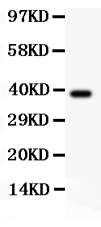
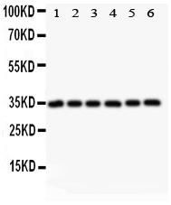
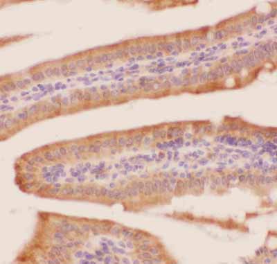
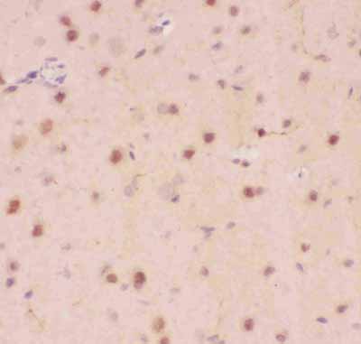
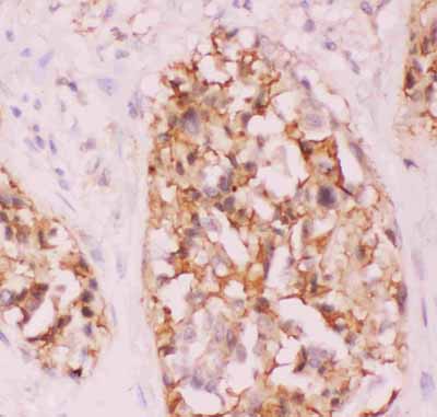
| Description | Rabbit IgG polyclonal antibody for Adapter molecule crk(CRK) detection. Tested with WB, IHC-P in Human;Mouse;Rat. |
| Reconstitution | Add 0.2ml of distilled water will yield a concentration of 500ug/ml. |
| Gene ID | 1398 |
|---|---|
| Other Names | Adapter molecule crk, Proto-oncogene c-Crk, p38, CRK |
| Calculated MW | 33831 MW KDa |
| Application Details | Immunohistochemistry(Paraffin-embedded Section), 0.5-1 µg/ml, Human, Mouse, Rat, By Heat Western blot, 0.1-0.5 µg/ml, Human, Mouse, Rat |
| Subcellular Localization | Cytoplasm . Cell membrane . Translocated to the plasma membrane upon cell adhesion. . |
| Protein Name | Adapter molecule crk |
| Contents | Each vial contains 5mg BSA, 0.9mg NaCl, 0.2mg Na2HPO4, 0.05mg NaN3. |
| Immunogen | E.coli-derived human Crk p38 recombinant protein (Position: A2-R246). Human Crk p38 shares 99% and 100% amino acid (aa) sequences identity with mouse and rat Crk p38, respectively. |
| Purification | Immunogen affinity purified. |
| Cross Reactivity | No cross reactivity with other proteins |
| Storage | At -20˚C for one year. After r˚Constitution, at 4˚C for one month. It˚Can also be aliquotted and stored frozen at -20˚C for a longer time.Avoid repeated freezing and thawing. |
| Sequence Similarities | Belongs to the CRK family. |
| Name | CRK |
|---|---|
| Function | Involved in cell branching and adhesion mediated by BCAR1- CRK-RAPGEF1 signaling and activation of RAP1. |
| Cellular Location | Cytoplasm. Cell membrane. Note=Translocated to the plasma membrane upon cell adhesion. |

Thousands of laboratories across the world have published research that depended on the performance of antibodies from Abcepta to advance their research. Check out links to articles that cite our products in major peer-reviewed journals, organized by research category.
info@abcepta.com, and receive a free "I Love Antibodies" mug.

Provided below are standard protocols that you may find useful for product applications.
Background
CRK, also known as p38 or CRKII, is a protein that in humans is encoded by the CRK gene. This gene is a member of an adapter protein family that binds to several tyrosine-phosphorylated proteins. It is mapped to 17p13.3. The CRK protein participates in the Reelin signaling cascade downstream of DAB1. The product of this gene has several SH2 and SH3 domains (src-homology domains) and is involved in several signaling pathways, recruiting cytoplasmic proteins in the vicinity of tyrosine kinase through SH2-phosphotyrosine interaction. The N-terminal SH2 domain of this protein functions as a positive regulator of transformation whereas the C-terminal SH3 domain functions as a negative regulator of transformation. Two alternative transcripts encoding different isoforms with distinct biological activity have been described.
If you have used an Abcepta product and would like to share how it has performed, please click on the "Submit Review" button and provide the requested information. Our staff will examine and post your review and contact you if needed.
If you have any additional inquiries please email technical services at tech@abcepta.com.
Ordering Information
Other Products
Shipping Information
