


 Foundational characteristics of cancer include proliferation, angiogenesis, migration, evasion of apoptosis, and cellular immortality. Find key markers for these cellular processes and antibodies to detect them.
 The SUMOplot™ Analysis Program predicts and scores sumoylation sites in your protein. SUMOylation is a post-translational modification involved in various cellular processes, such as nuclear-cytosolic transport, transcriptional regulation, apoptosis, protein stability, response to stress, and progression through the cell cycle.
 The Autophagy Receptor Motif Plotter predicts and scores autophagy receptor binding sites in your protein. Identifying proteins connected to this pathway is critical to understanding the role of autophagy in physiological as well as pathological processes such as development, differentiation, neurodegenerative diseases, stress, infection, and cancer.
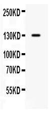
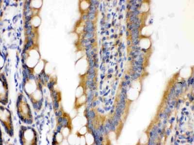
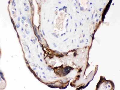
Anti-CDCP1 Picoband Antibody

- SPECIFICATION
- CITATIONS
- PROTOCOLS
- BACKGROUND

Application
| WB, IHC-P |
|---|---|
| Primary Accession | Q9H5V8 |
| Host | Rabbit |
| Reactivity | Human, Mouse, Rat |
| Clonality | Polyclonal |
| Format | Lyophilized |
| Description | Rabbit IgG polyclonal antibody for CUB domain-containing protein 1(CDCP1) detection. Tested with WB, IHC-P in Human;Mouse;Rat. |
| Reconstitution | Add 0.2ml of distilled water will yield a concentration of 500ug/ml. |
| Gene ID | 64866 |
|---|---|
| Other Names | CUB domain-containing protein 1, Membrane glycoprotein gp140, Subtractive immunization M plus HEp3-associated 135 kDa protein, SIMA135, Transmembrane and associated with src kinases, CD318, CDCP1, TRASK |
| Calculated MW | 92932 MW KDa |
| Application Details | Immunohistochemistry(Paraffin-embedded Section), 0.5-1 µg/ml, Human, Mouse, Rat, By Heat Western blot, 0.1-0.5 µg/ml, Human |
| Subcellular Localization | Isoform 1: Cell membrane ; Single-pass membrane protein . Shedding may also lead to a soluble peptide. |
| Tissue Specificity | Highly expressed in mitotic cells with low expression during interphase. Detected at highest levels in skeletal muscle and colon with lower levels in kidney, small intestine, placenta and lung. Up-regulated in a number of human tumor cell lines, as well as in colorectal cancer, breast carcinoma and lung cancer. Also expressed in cells with phenotypes reminiscent of mesenchymal stem cells and neural stem cells. . |
| Protein Name | CUB domain-containing protein 1 |
| Contents | Each vial contains 5mg BSA, 0.9mg NaCl, 0.2mg Na2HPO4, 0.05mg NaN3. |
| Immunogen | E. coli-derived human CDCP1 recombinant protein (Position: R582-T667). Human CDCP1 shares 84.5% amino acid (aa) sequence identity with mouse CDCP1. |
| Purification | Immunogen affinity purified. |
| Cross Reactivity | No cross reactivity with other proteins |
| Storage | At -20˚C for one year. After r˚Constitution, at 4˚C for one month. It˚Can also be aliquotted and stored frozen at -20˚C for a longer time.Avoid repeated freezing and thawing. |
| Name | CDCP1 |
|---|---|
| Synonyms | TRASK |
| Function | May be involved in cell adhesion and cell matrix association. May play a role in the regulation of anchorage versus migration or proliferation versus differentiation via its phosphorylation. May be a novel marker for leukemia diagnosis and for immature hematopoietic stem cell subsets. Belongs to the tetraspanin web involved in tumor progression and metastasis. |
| Cellular Location | [Isoform 1]: Cell membrane; Single- pass membrane protein. Note=Shedding may also lead to a soluble peptide |
| Tissue Location | Highly expressed in mitotic cells with low expression during interphase. Detected at highest levels in skeletal muscle and colon with lower levels in kidney, small intestine, placenta and lung. Up-regulated in a number of human tumor cell lines, as well as in colorectal cancer, breast carcinoma and lung cancer. Also expressed in cells with phenotypes reminiscent of mesenchymal stem cells and neural stem cells. |

Thousands of laboratories across the world have published research that depended on the performance of antibodies from Abcepta to advance their research. Check out links to articles that cite our products in major peer-reviewed journals, organized by research category.
info@abcepta.com, and receive a free "I Love Antibodies" mug.

Provided below are standard protocols that you may find useful for product applications.
Background
CUB domain-containing protein 1 (CDCP1) is a protein that in humans is encoded by the CDCP1 gene. It has also been designated as CD318 (cluster of differentiation 318) and Trask (Transmembrane and associated with src kinases). CDCP1/Trask is a 140 kD transmembrane glycoprotein with a large extracellular domain (ECD) containing two CUB domains, and a smaller intracellular domain (ICD) containing five tyrosines. The tyrosine phosphorylation of Trask is tightly regulated and reciprocally linked with the state of cell adhesion. The tyrosine phosphorylation of CDCP1 in cultured cells occurs when cells are induced to detach by trypsin or EDTA, or seen spontaneously during mitotic detachment. The overexpression of CDCP1 leads to the loss of cell adhesion and a detached phenotype. CDCP1 is widely expressed in human epithelial tissues, but its phosphorylation is only seen in mitotically detached or shedding cells, consistent with its role in the negative regulation of cell adhesion.
If you have used an Abcepta product and would like to share how it has performed, please click on the "Submit Review" button and provide the requested information. Our staff will examine and post your review and contact you if needed.
If you have any additional inquiries please email technical services at tech@abcepta.com.
Ordering Information
Other Products
Shipping Information
