


 Foundational characteristics of cancer include proliferation, angiogenesis, migration, evasion of apoptosis, and cellular immortality. Find key markers for these cellular processes and antibodies to detect them.
 The SUMOplot™ Analysis Program predicts and scores sumoylation sites in your protein. SUMOylation is a post-translational modification involved in various cellular processes, such as nuclear-cytosolic transport, transcriptional regulation, apoptosis, protein stability, response to stress, and progression through the cell cycle.
 The Autophagy Receptor Motif Plotter predicts and scores autophagy receptor binding sites in your protein. Identifying proteins connected to this pathway is critical to understanding the role of autophagy in physiological as well as pathological processes such as development, differentiation, neurodegenerative diseases, stress, infection, and cancer.
> home > Products > Primary Antibodies > Neuroscience > Anti-NMDAR1 GRIN1 Rabbit Monoclonal Antibody
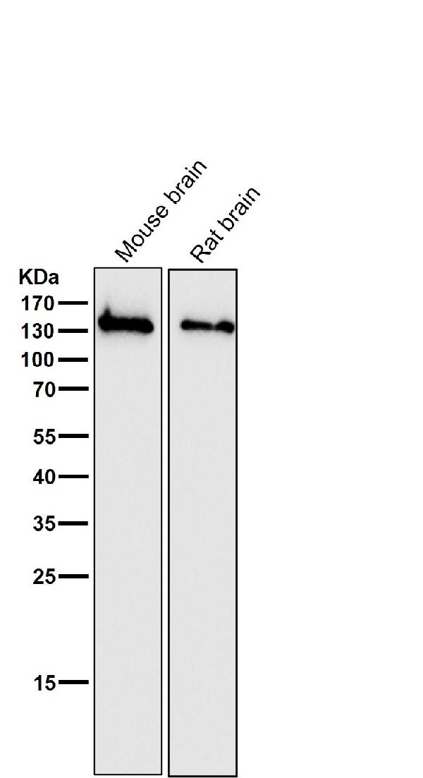
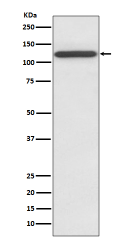
Anti-NMDAR1 GRIN1 Rabbit Monoclonal Antibody
- SPECIFICATION
- CITATIONS
- PROTOCOLS
- BACKGROUND

Application
| WB |
|---|---|
| Primary Accession | Q05586 |
| Host | Rabbit |
| Isotype | Rabbit IgG |
| Reactivity | Rat, Human, Mouse |
| Clonality | Monoclonal |
| Format | Liquid |
| Description | Anti-NMDAR1 GRIN1 Rabbit Monoclonal Antibody . Tested in WB application. This antibody reacts with Human, Mouse, Rat. |
| Gene ID | 2902 |
|---|---|
| Other Names | Glutamate receptor ionotropic, NMDA 1, GluN1, Glutamate [NMDA] receptor subunit zeta-1, N-methyl-D-aspartate receptor subunit NR1, NMD-R1, GRIN1, NMDAR1 |
| Calculated MW | 105373 MW KDa |
| Application Details | WB 1:500-1:2000 |
| Subcellular Localization | Cell membrane ; Multi-pass membrane protein. Cell junction, synapse, postsynaptic cell membrane. Cell junction, synapse, postsynaptic cell membrane, postsynaptic density. Enriched in postsynaptic plasma membrane and postsynaptic densities.. |
| Contents | Rabbit IgG in phosphate buffered saline, pH 7.4, 150mM NaCl, 0.02% sodium azide and 50% glycerol, 0.4-0.5mg/ml BSA. |
| Clone Names | Clone: AAHF-7 |
| Immunogen | A synthesized peptide derived from human NMDAR1 |
| Purification | Affinity-chromatography |
| Storage | Store at -20°C for one year. For short term storage and frequent use, store at 4°C for up to one month. Avoid repeated freeze-thaw cycles. |
| Name | GRIN1 (HGNC:4584) |
|---|---|
| Function | Component of N-methyl-D-aspartate (NMDA) receptors (NMDARs) that function as heterotetrameric, ligand-gated cation channels with high calcium permeability and voltage-dependent block by Mg(2+) (PubMed:21376300, PubMed:26875626, PubMed:26919761, PubMed:28126851, PubMed:28228639, PubMed:36959261, PubMed:7679115, PubMed:7681588, PubMed:7685113). NMDARs participate in synaptic plasticity for learning and memory formation by contributing to the long-term potentiation (LTP) (PubMed:26875626). Channel activation requires binding of the neurotransmitter L-glutamate to the GluN2 subunit, glycine or D-serine binding to the GluN1 subunit, plus membrane depolarization to eliminate channel inhibition by Mg(2+) (PubMed:21376300, PubMed:26875626, PubMed:26919761, PubMed:27164704, PubMed:28095420, PubMed:28105280, PubMed:28126851, PubMed:28228639, PubMed:36959261, PubMed:38538865, PubMed:7679115, PubMed:7681588, PubMed:7685113). NMDARs mediate simultaneously the potasium efflux and the influx of calcium and sodium (By similarity). Each GluN2 or GluN3 subunit confers differential attributes to channel properties, including activation, deactivation and desensitization kinetics, pH sensitivity, Ca2(+) permeability, and binding to allosteric modulators (PubMed:26875626, PubMed:26919761, PubMed:36309015, PubMed:38598639). |
| Cellular Location | Cell membrane; Multi-pass membrane protein {ECO:0000250|UniProtKB:P35439}. Postsynaptic cell membrane {ECO:0000250|UniProtKB:P35438}. Postsynaptic density membrane {ECO:0000250|UniProtKB:P35439}. Synaptic cell membrane {ECO:0000250|UniProtKB:P35438}. Note=Synaptic cell membrane targeting is dependent of GRIN2B/GluN2B subunit (By similarity). Association with GRIN3A occurs in the endoplasmic reticulum (By similarity) {ECO:0000250, ECO:0000250|UniProtKB:P35438, ECO:0000250|UniProtKB:P35439} |
Research Areas
Citations (0)

Thousands of laboratories across the world have published research that depended on the performance of antibodies from Abcepta to advance their research. Check out links to articles that cite our products in major peer-reviewed journals, organized by research category.
Submit your citation using an Abcepta antibody to
info@abcepta.com, and receive a free "I Love Antibodies" mug.
info@abcepta.com, and receive a free "I Love Antibodies" mug.

Application Protocols
Provided below are standard protocols that you may find useful for product applications.
Abcepta welcomes feedback from its customers.
If you have used an Abcepta product and would like to share how it has performed, please click on the "Submit Review" button and provide the requested information. Our staff will examine and post your review and contact you if needed.
If you have any additional inquiries please email technical services at tech@abcepta.com.
$ 370.00
Cat# ABO13873
Ordering Information
United States
AlbaniaAustraliaAustriaBelgiumBosnia & HerzegovinaBrazilBulgariaCanadaCentral AmericaChinaCroatiaCyprusCzech RepublicDenmarkEstoniaFinlandFranceGermanyGreeceHong KongHungaryIcelandIndiaIndonesiaIrelandIsraelItalyJapanLatviaLithuaniaLuxembourgMacedoniaMalaysiaMaltaMexicoNetherlandsNew ZealandNorwayPakistanPolandPortugalRomaniaSerbiaSingaporeSlovakiaSloveniaSouth AfricaSouth KoreaSpainSwedenSwitzerlandTaiwanTurkeyUnited KingdomUnited StatesVietnamWorldwideOthers
USA Headquarters
(888) 735-7227 / (858) 622-0099 or (858) 875-1900
Other Products
Shipping Information
Domestic orders (in stock items)
Shipped out the same day. Orders placed after 1 PM (PST) will ship out the next business day.
International orders
Contact your local distributors
