


 Foundational characteristics of cancer include proliferation, angiogenesis, migration, evasion of apoptosis, and cellular immortality. Find key markers for these cellular processes and antibodies to detect them.
 The SUMOplot™ Analysis Program predicts and scores sumoylation sites in your protein. SUMOylation is a post-translational modification involved in various cellular processes, such as nuclear-cytosolic transport, transcriptional regulation, apoptosis, protein stability, response to stress, and progression through the cell cycle.
 The Autophagy Receptor Motif Plotter predicts and scores autophagy receptor binding sites in your protein. Identifying proteins connected to this pathway is critical to understanding the role of autophagy in physiological as well as pathological processes such as development, differentiation, neurodegenerative diseases, stress, infection, and cancer.
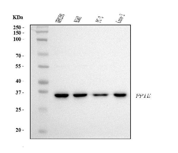
Anti-Cyclophilin E/PPIE Antibody Picoband™ (monoclonal, 7F2)
- SPECIFICATION
- CITATIONS
- PROTOCOLS
- BACKGROUND

Application
| WB, IHC, IF, ICC, FC |
|---|---|
| Primary Accession | Q9UNP9 |
| Host | Mouse |
| Isotype | Mouse IgG2a |
| Reactivity | Rat, Human, Mouse |
| Clonality | Monoclonal |
| Format | Lyophilized |
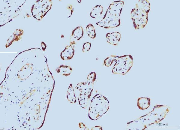
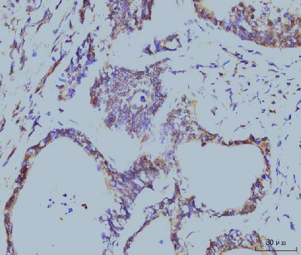
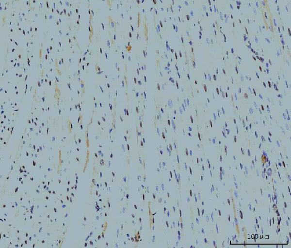
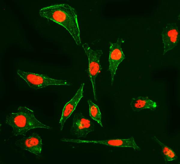
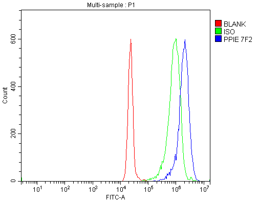
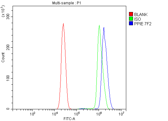
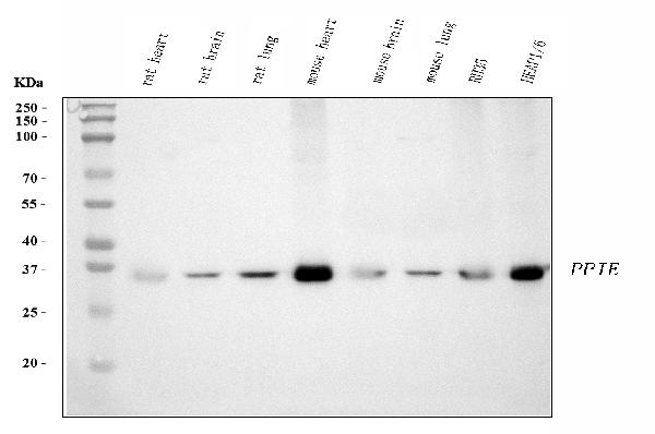
| Description | Anti-Cyclophilin E/PPIE Antibody Picoband™ (monoclonal, 7F2) . Tested in Flow Cytometry, IF, IHC, ICC, WB applications. This antibody reacts with Human, Mouse, Rat. |
| Reconstitution | Adding 0.2 ml of distilled water will yield a concentration of 500 µg/ml. |
| Gene ID | 10450 |
|---|---|
| Other Names | Peptidyl-prolyl cis-trans isomerase E, PPIase E, 5.2.1.8, Cyclophilin E, Cyclophilin-33, Rotamase E, PPIE, CYP33 {ECO:0000303|PubMed:8977107} |
| Calculated MW | 35 kDa |
| Application Details | Western blot, 0.25-0.5 µg/ml, Human, Mouse, Rat Immunohistochemistry(Paraffin-embedded Section), 2-5 µg/ml, Human, Rat Immunocytochemistry/Immunofluorescence, 5 µg/ml, Human Flow Cytometry, 1-3 µg/1x10^6 cells, Human |
| Contents | Each vial contains 4 mg Trehalose, 0.9 mg NaCl and 0.2 mg Na2HPO4. |
| Clone Names | Clone: 7F2 |
| Immunogen | E.coli-derived human Cyclophilin E/PPIE recombinant protein (Position: M1-V301). |
| Purification | Immunogen affinity purified. |
| Storage | At -20°C for one year from date of receipt. After reconstitution, at 4°C for one month. It can also be aliquotted and stored frozen at -20°C for six months. Avoid repeated freezing and thawing. |
| Name | PPIE |
|---|---|
| Synonyms | CYP33 {ECO:0000303|PubMed:8977107} |
| Function | Involved in pre-mRNA splicing as component of the spliceosome (PubMed:11991638, PubMed:28076346). Combines RNA-binding and PPIase activities (PubMed:18258190, PubMed:20460131, PubMed:20677832, PubMed:8977107). Binds mRNA and has a preference for single-stranded RNA molecules with poly-A and poly-U stretches, suggesting it binds to the poly(A)-region in the 3'-UTR of mRNA molecules (PubMed:18258190, PubMed:20460131, PubMed:8977107). Catalyzes the cis-trans isomerization of proline imidic peptide bonds in proteins (PubMed:18258190, PubMed:20541251, PubMed:20677832, PubMed:8977107). Inhibits KMT2A activity; this requires proline isomerase activity (PubMed:20460131, PubMed:20541251, PubMed:20677832). |
| Cellular Location | Nucleus |
| Tissue Location | Found in all the examined tissues including heart, brain, placenta, lung, liver, skeletal muscle, kidney and pancreas |

Thousands of laboratories across the world have published research that depended on the performance of antibodies from Abcepta to advance their research. Check out links to articles that cite our products in major peer-reviewed journals, organized by research category.
info@abcepta.com, and receive a free "I Love Antibodies" mug.

Provided below are standard protocols that you may find useful for product applications.
Background
Peptidylprolyl isomerase E (cyclophilin E), also known as PPIE, is an enzyme which in humans is encoded by the PPIE gene on chromosome 1. The protein encoded by this gene is a member of the peptidyl-prolyl cis-trans isomerase (PPIase) family. PPIases catalyze the cis-trans isomerization of proline imidic peptide bonds in oligopeptides and accelerate the folding of proteins. This protein contains a highly conserved cyclophilin (CYP) domain as well as an RNA-binding domain. It was shown to possess PPIase and protein folding activities, and it also exhibits RNA-binding activity. Alternative splicing results in multiple transcript variants. A related pseudogene, which is also located on chromosome 1, has been identified.
If you have used an Abcepta product and would like to share how it has performed, please click on the "Submit Review" button and provide the requested information. Our staff will examine and post your review and contact you if needed.
If you have any additional inquiries please email technical services at tech@abcepta.com.
Ordering Information
Other Products
Shipping Information
