


 Foundational characteristics of cancer include proliferation, angiogenesis, migration, evasion of apoptosis, and cellular immortality. Find key markers for these cellular processes and antibodies to detect them.
 The SUMOplot™ Analysis Program predicts and scores sumoylation sites in your protein. SUMOylation is a post-translational modification involved in various cellular processes, such as nuclear-cytosolic transport, transcriptional regulation, apoptosis, protein stability, response to stress, and progression through the cell cycle.
 The Autophagy Receptor Motif Plotter predicts and scores autophagy receptor binding sites in your protein. Identifying proteins connected to this pathway is critical to understanding the role of autophagy in physiological as well as pathological processes such as development, differentiation, neurodegenerative diseases, stress, infection, and cancer.
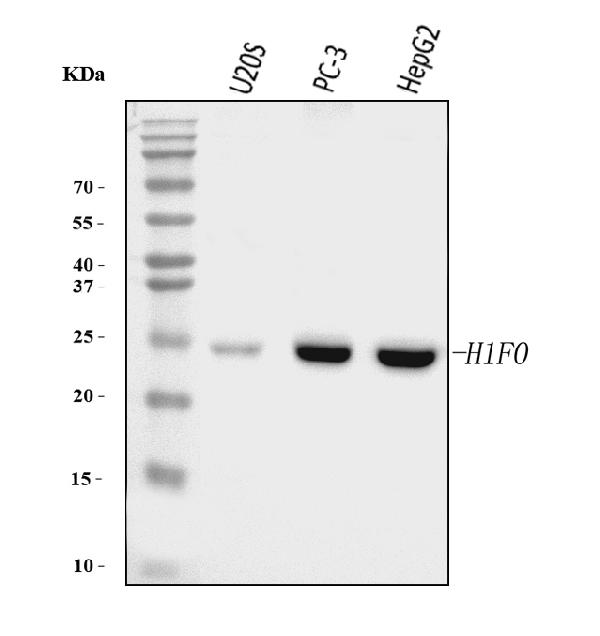
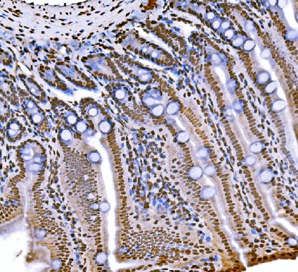
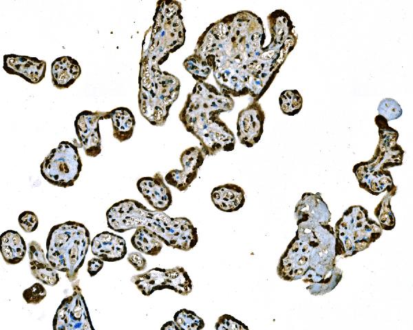
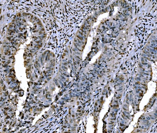
Anti-Histone H1.0/H1F0 Antibody Picoband™ (monoclonal, 5I3E6)
- SPECIFICATION
- CITATIONS
- PROTOCOLS
- BACKGROUND

Application
| WB, IHC, FC |
|---|---|
| Primary Accession | P07305 |
| Host | Mouse |
| Isotype | Mouse IgG2b |
| Reactivity | Human, Mouse |
| Clonality | Monoclonal |
| Format | Lyophilized |
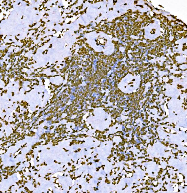
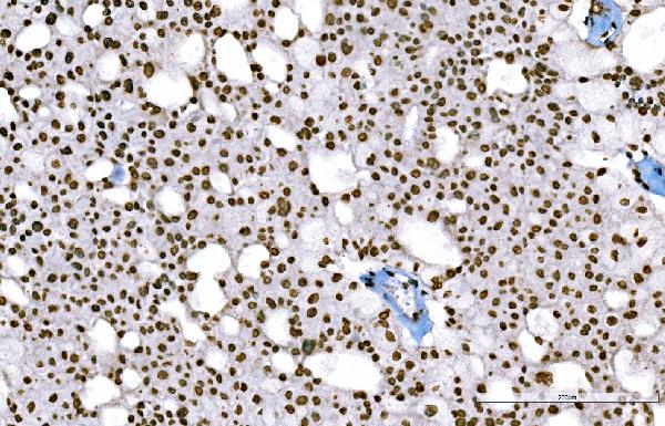
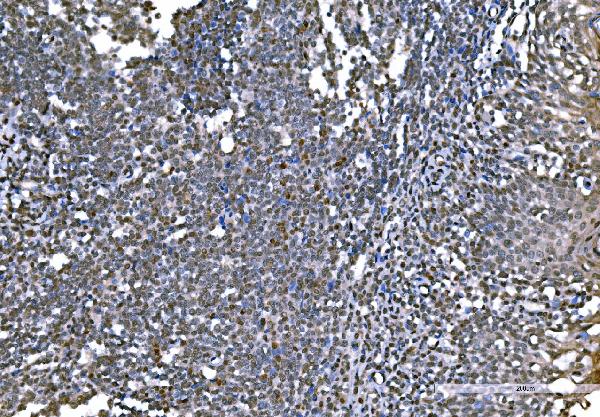
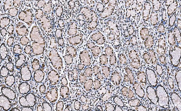
| Description | Anti-Histone H1.0/H1F0 Antibody Picoband™ (monoclonal, 5I3E6) . Tested in Flow Cytometry, IHC, WB applications. This antibody reacts with Human, Mouse. |
| Reconstitution | Adding 0.2 ml of distilled water will yield a concentration of 500 µg/ml. |
| Gene ID | 3005 |
|---|---|
| Other Names | Histone H1.0, Histone H1', Histone H1(0), Histone H1.0, N-terminally processed, H1-0 (HGNC:4714) |
| Calculated MW | 24 kDa |
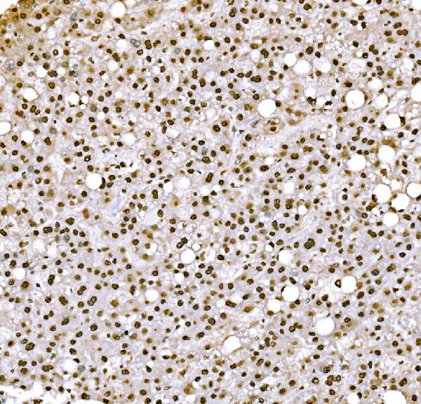
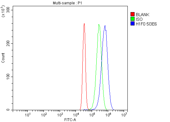
| Application Details | Western blot, 0.25-0.5 µg/ml, Human Immunohistochemistry(Paraffin-embedded Section), 2-5 µg/ml, Human, Mouse Flow Cytometry, 1-3 µg/1x10^6 cells, Human |
| Contents | Each vial contains 4 mg Trehalose, 0.9 mg NaCl and 0.2 mg Na2HPO4. |
| Clone Names | Clone: 5I3E6 |
| Immunogen | E.coli-derived human Histone H1.0/H1F0 recombinant protein (Position: K20-K159). |
| Purification | Immunogen affinity purified. |
| Storage | At -20°C for one year from date of receipt. After reconstitution, at 4°C for one month. It can also be aliquotted and stored frozen at -20°C for six months. Avoid repeated freezing and thawing. |
| Name | H1-0 (HGNC:4714) |
|---|---|
| Function | Histones H1 are necessary for the condensation of nucleosome chains into higher-order structures. The histones H1.0 are found in cells that are in terminal stages of differentiation or that have low rates of cell division. |
| Cellular Location | Nucleus {ECO:0000255|PROSITE-ProRule:PRU00837, ECO:0000269|PubMed:18993075}. Chromosome {ECO:0000255|PROSITE- ProRule:PRU00837, ECO:0000269|PubMed:18993075}. Note=The RNA edited version has been localized to nuclear speckles. During mitosis, it appears in the vicinity of condensed chromosomes |

Thousands of laboratories across the world have published research that depended on the performance of antibodies from Abcepta to advance their research. Check out links to articles that cite our products in major peer-reviewed journals, organized by research category.
info@abcepta.com, and receive a free "I Love Antibodies" mug.

Provided below are standard protocols that you may find useful for product applications.
Background
H1 histone family, member 0is a member of thehistonefamily of nuclearproteinswhich are a component ofchromatin. In humans, this protein is encoded by theH1F0gene. It is mapped to 22q13.1. Histones are basic nuclear proteins that are responsible for the nucleosome structure of the chromosomal fiber in eukaryotes. Nucleosomes consist of approximately 146 bp of DNA wrapped around a histone octamer composed of pairs of each of the four core histones (H2A, H2B, H3, and H4). The chromatin fiber is further compacted through the interaction of a linker histone, H1, with the DNA between the nucleosomes to form higher order chromatin structures. This gene is intronless and encodes a replication-independent histone that is a member of the histone H1 family.
If you have used an Abcepta product and would like to share how it has performed, please click on the "Submit Review" button and provide the requested information. Our staff will examine and post your review and contact you if needed.
If you have any additional inquiries please email technical services at tech@abcepta.com.
Ordering Information
Other Products
Shipping Information
