


 Foundational characteristics of cancer include proliferation, angiogenesis, migration, evasion of apoptosis, and cellular immortality. Find key markers for these cellular processes and antibodies to detect them.
 The SUMOplot™ Analysis Program predicts and scores sumoylation sites in your protein. SUMOylation is a post-translational modification involved in various cellular processes, such as nuclear-cytosolic transport, transcriptional regulation, apoptosis, protein stability, response to stress, and progression through the cell cycle.
 The Autophagy Receptor Motif Plotter predicts and scores autophagy receptor binding sites in your protein. Identifying proteins connected to this pathway is critical to understanding the role of autophagy in physiological as well as pathological processes such as development, differentiation, neurodegenerative diseases, stress, infection, and cancer.
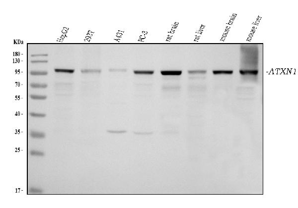
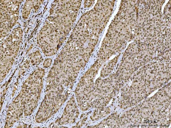
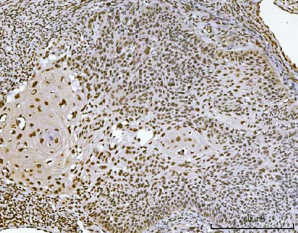
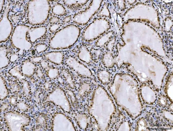
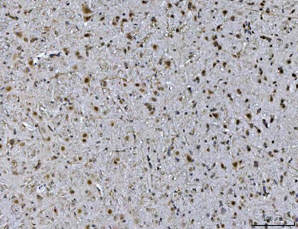
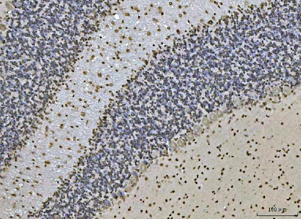
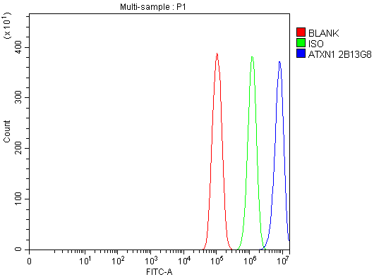
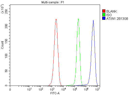
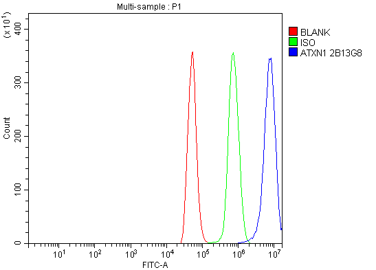
Anti-Ataxin 1 Antibody Picoband™ (monoclonal, 2B13G8)
- SPECIFICATION
- CITATIONS
- PROTOCOLS
- BACKGROUND

Application
| WB, IHC, FC |
|---|---|
| Primary Accession | P54253 |
| Host | Mouse |
| Isotype | Mouse IgG2b |
| Reactivity | Rat, Human, Mouse |
| Clonality | Monoclonal |
| Format | Lyophilized |
| Description | Anti-Ataxin 1 Antibody Picoband™ (monoclonal, 2B13G8) . Tested in Flow Cytometry, IHC, WB applications. This antibody reacts with Human, Mouse, Rat. |
| Reconstitution | Adding 0.2 ml of distilled water will yield a concentration of 500 µg/ml. |
| Gene ID | 6310 |
|---|---|
| Other Names | Ataxin-1, Spinocerebellar ataxia type 1 protein, ATXN1, ATX1, SCA1 |
| Calculated MW | 105 kDa |
| Application Details | Western blot, 0.25-0.5 µg/ml, Human, Mouse, Rat Immunohistochemistry(Paraffin-embedded Section), 2-5 µg/ml, Human, Mouse, Rat Flow Cytometry, 1-3 µg/1x10^6 cells, Human, Mouse, Rat |
| Contents | Each vial contains 4 mg Trehalose, 0.9 mg NaCl and 0.2 mg Na2HPO4. |
| Clone Names | Clone: 2B13G8 |
| Immunogen | A synthetic peptide corresponding to a sequence at the C-terminus of human Ataxin 1, different from the related mouse and rat sequences by one amino acid. |
| Purification | Immunogen affinity purified. |
| Storage | At -20°C for one year from date of receipt. After reconstitution, at 4°C for one month. It can also be aliquotted and stored frozen at -20°C for six months. Avoid repeated freezing and thawing. |
| Name | ATXN1 |
|---|---|
| Synonyms | ATX1, SCA1 |
| Function | Chromatin-binding factor that repress Notch signaling in the absence of Notch intracellular domain by acting as a CBF1 corepressor. Binds to the HEY promoter and might assist, along with NCOR2, RBPJ- mediated repression. Binds RNA in vitro. May be involved in RNA metabolism (PubMed:21475249). In concert with CIC and ATXN1L, involved in brain development (By similarity). |
| Cellular Location | Cytoplasm. Nucleus Note=Colocalizes with USP7 in the nucleus |
| Tissue Location | Widely expressed throughout the body. |

Thousands of laboratories across the world have published research that depended on the performance of antibodies from Abcepta to advance their research. Check out links to articles that cite our products in major peer-reviewed journals, organized by research category.
info@abcepta.com, and receive a free "I Love Antibodies" mug.

Provided below are standard protocols that you may find useful for product applications.
Background
Ataxin-1 is a protein that in humans is encoded by the ATXN1 gene. The ATXN1 gene had been mapped to 6p23 by in situ hybridization. Ataxin-1 (ATXN1), a causative factor for spinocerebellar ataxia type 1 (SCA1), and the related Brother of ATXN1 (BOAT1) are human proteins involved in transcriptional repression. ATXN1 and BOAT1 might participate in several Notch-controlled developmental and pathological processes.
If you have used an Abcepta product and would like to share how it has performed, please click on the "Submit Review" button and provide the requested information. Our staff will examine and post your review and contact you if needed.
If you have any additional inquiries please email technical services at tech@abcepta.com.
Ordering Information
Other Products
Shipping Information
