


 Foundational characteristics of cancer include proliferation, angiogenesis, migration, evasion of apoptosis, and cellular immortality. Find key markers for these cellular processes and antibodies to detect them.
 The SUMOplot™ Analysis Program predicts and scores sumoylation sites in your protein. SUMOylation is a post-translational modification involved in various cellular processes, such as nuclear-cytosolic transport, transcriptional regulation, apoptosis, protein stability, response to stress, and progression through the cell cycle.
 The Autophagy Receptor Motif Plotter predicts and scores autophagy receptor binding sites in your protein. Identifying proteins connected to this pathway is critical to understanding the role of autophagy in physiological as well as pathological processes such as development, differentiation, neurodegenerative diseases, stress, infection, and cancer.
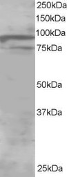
Goat Anti-ORP11 / OSBPL11 Antibody
Peptide-affinity purified goat antibody
- SPECIFICATION
- CITATIONS
- PROTOCOLS
- BACKGROUND

Application
| WB, E |
|---|---|
| Primary Accession | Q9BXB4 |
| Other Accession | NP_073613, 114885 |
| Reactivity | Human |
| Predicted | Mouse, Rat, Dog |
| Host | Goat |
| Clonality | Polyclonal |
| Concentration | 100ug/200ul |
| Isotype | IgG |
| Calculated MW | 83643 Da |
| Gene ID | 114885 |
|---|---|
| Other Names | Oxysterol-binding protein-related protein 11, ORP-11, OSBP-related protein 11, OSBPL11, ORP11, OSBP12 |
| Dilution | WB~~1:1000 E~~N/A |
| Format | 0.5 mg IgG/ml in Tris saline (20mM Tris pH7.3, 150mM NaCl), 0.02% sodium azide, with 0.5% bovine serum albumin |
| Storage | Maintain refrigerated at 2-8°C for up to 6 months. For long term storage store at -20°C in small aliquots to prevent freeze-thaw cycles. |
| Precautions | Goat Anti-ORP11 / OSBPL11 Antibody is for research use only and not for use in diagnostic or therapeutic procedures. |
| Name | OSBPL11 |
|---|---|
| Synonyms | ORP11, OSBP12 |
| Function | Plays a role in regulating ADIPOQ and FABP4 levels in differentiating adipocytes and is also involved in regulation of adipocyte triglyceride storage (PubMed:23028956). Weakly binds 25- hydroxycholesterol (PubMed:17428193). Interacts with OSBPL9 to function as lipid transfer proteins (PubMed:39106189). Together they form a heterodimer that localizes at the ER-trans-Golgi membrane contact sites, and exchanges phosphatidylserine (1,2-diacyl-sn-glycero-3- phospho-L-serine, PS) for phosphatidylinositol-4-phosphate (1,2-diacyl- sn-glycero-3-phospho-(1D-myo-inositol 4-phosphate), PI(4)P) between the two organelles, a step that is critical for sphingomyelin synthesis in the Golgi complex (PubMed:39106189). |
| Cellular Location | Late endosome membrane. Golgi apparatus, trans-Golgi network membrane. Note=Localizes at the Golgi- late endosome interface |
| Tissue Location | Present at highest levels in ovary, testis, kidney, liver, stomach, brain, and adipose tissue. Strong expression (at protein level) in epithelial cells of kidney tubules, testicular tubules, caecum, and skin (PubMed:20599956). Present at low levels in subcutaneous and visceral adipose tissue (at protein level) (PubMed:23028956). |

Thousands of laboratories across the world have published research that depended on the performance of antibodies from Abcepta to advance their research. Check out links to articles that cite our products in major peer-reviewed journals, organized by research category.
info@abcepta.com, and receive a free "I Love Antibodies" mug.

Provided below are standard protocols that you may find useful for product applications.
Background
This gene encodes a member of the oxysterol-binding protein (OSBP) family, a group of intracellular lipid receptors. Like most members, the encoded protein contains an N-terminal pleckstrin homology domain and a highly conserved C-terminal OSBP-like sterol-binding domain.
References
Variation at the NFATC2 Locus Increases the Risk of Thiazolinedinedione-Induced Edema in the Diabetes REduction Assessment with ramipril and rosiglitazone Medication (DREAM) Study. Bailey SD, et al. Diabetes Care, 2010 Jul 13. PMID 20628086.
Gene-centric association signals for lipids and apolipoproteins identified via the HumanCVD BeadChip. Talmud PJ, et al. Am J Hum Genet, 2009 Nov. PMID 19913121.
Defining the human deubiquitinating enzyme interaction landscape. Sowa ME, et al. Cell, 2009 Jul 23. PMID 19615732.
Association of OSBPL11 gene polymorphisms with cardiovascular disease risk factors in obesity. Bouchard L, et al. Obesity (Silver Spring), 2009 Jul. PMID 19325544.
Global, in vivo, and site-specific phosphorylation dynamics in signaling networks. Olsen JV, et al. Cell, 2006 Nov 3. PMID 17081983.
If you have used an Abcepta product and would like to share how it has performed, please click on the "Submit Review" button and provide the requested information. Our staff will examine and post your review and contact you if needed.
If you have any additional inquiries please email technical services at tech@abcepta.com.
Ordering Information
Other Products
Shipping Information
