


 Foundational characteristics of cancer include proliferation, angiogenesis, migration, evasion of apoptosis, and cellular immortality. Find key markers for these cellular processes and antibodies to detect them.
 The SUMOplot™ Analysis Program predicts and scores sumoylation sites in your protein. SUMOylation is a post-translational modification involved in various cellular processes, such as nuclear-cytosolic transport, transcriptional regulation, apoptosis, protein stability, response to stress, and progression through the cell cycle.
 The Autophagy Receptor Motif Plotter predicts and scores autophagy receptor binding sites in your protein. Identifying proteins connected to this pathway is critical to understanding the role of autophagy in physiological as well as pathological processes such as development, differentiation, neurodegenerative diseases, stress, infection, and cancer.
VPS41 Antibody (internal region)
Peptide-affinity purified goat antibody
- SPECIFICATION
- CITATIONS
- PROTOCOLS
- BACKGROUND

Application
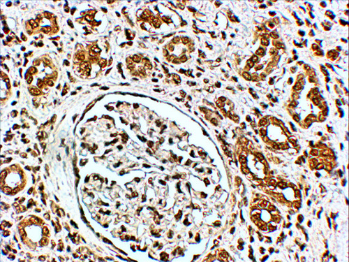
| IHC, E |
|---|---|
| Primary Accession | P49754 |
| Other Accession | NP_055211.2, NP_542198.2, 27072 |
| Reactivity | Human |
| Predicted | Mouse, Rat, Dog |
| Host | Goat |
| Clonality | Polyclonal |
| Concentration | 0.5 mg/ml |
| Isotype | IgG |
| Calculated MW | 98566 Da |
| Gene ID | 27072 |
|---|---|
| Other Names | Vacuolar protein sorting-associated protein 41 homolog, S53, VPS41 |
| Dilution | IHC~~1:100~500 E~~N/A |
| Format | 0.5 mg/ml in Tris saline, 0.02% sodium azide, pH7.3 with 0.5% bovine serum albumin |
| Storage | Maintain refrigerated at 2-8°C for up to 6 months. For long term storage store at -20°C in small aliquots to prevent freeze-thaw cycles. |
| Precautions | VPS41 Antibody (internal region) is for research use only and not for use in diagnostic or therapeutic procedures. |
| Name | VPS41 |
|---|---|
| Function | Plays a role in vesicle-mediated protein trafficking to lysosomal compartments including the endocytic membrane transport and autophagic pathways. Acts as a component of the HOPS endosomal tethering complex. This complex is proposed to be involved in the Rab5- to-Rab7 endosome conversion probably implicating MON1A/B, and via binding SNAREs and SNARE complexes to mediate tethering and docking events during SNARE-mediated membrane fusion. The HOPS complex is proposed to be recruited to Rab7 on the late endosomal membrane and to regulate late endocytic, phagocytic and autophagic traffic towards lysosomes (PubMed:23351085, PubMed:33851776). Involved in homotypic vesicle fusions between late endosomes and in heterotypic fusions between late endosomes and lysosomes implicated in degradation of endocytosed cargo (PubMed:23167963, PubMed:25445562, PubMed:25908847, PubMed:9159129). Required for fusion of autophagosomes with lysosomes (PubMed:25783203, PubMed:37821429). Links the HOPS complex to endosomal Rab7 via its association with RILP and to lysosomal membranes via its association with ARL8B, suggesting that these interactions may bring the compartments to close proximity for fusion (PubMed:21802320, PubMed:25445562, PubMed:25908847). Involved in the direct trans-Golgi network to late endosomes transport of lysosomal membrane proteins independently of HOPS (PubMed:23322049). Involved in sorting to the regulated secretory pathway presumably implicating the AP-3 adapter complex (By similarity). May play a role in HOPS-independent function in the regulated secretory pathway (PubMed:24210660). |
| Cellular Location | Endosome membrane; Peripheral membrane protein. Late endosome membrane; Peripheral membrane protein. Early endosome membrane; Peripheral membrane protein. Lysosome membrane; Peripheral membrane protein. Golgi apparatus, trans- Golgi network. Cytoplasmic vesicle, clathrin-coated vesicle. Cytoplasm, cytosol |
| Tissue Location | Expressed in cerebral cortex and cerebellum. Highly expressed in Purkinje cells. |

Thousands of laboratories across the world have published research that depended on the performance of antibodies from Abcepta to advance their research. Check out links to articles that cite our products in major peer-reviewed journals, organized by research category.
info@abcepta.com, and receive a free "I Love Antibodies" mug.

Provided below are standard protocols that you may find useful for product applications.
Background
This antibody is expected to recognise both reported isoforms (NP_055211.2 and NP_542198.2)
References
hVPS41 is expressed in multiple isoforms and can associate with vesicles through a RING-H2 finger motif. McVey Ward D, Radisky D, Scullion MA, Tuttle MS, Vaughn M, Kaplan J. Exp Cell Res. 2001 Jul 1;267(1):126-34. PMID: 11412045
If you have used an Abcepta product and would like to share how it has performed, please click on the "Submit Review" button and provide the requested information. Our staff will examine and post your review and contact you if needed.
If you have any additional inquiries please email technical services at tech@abcepta.com.
Ordering Information
Other Products
Shipping Information
