


 Foundational characteristics of cancer include proliferation, angiogenesis, migration, evasion of apoptosis, and cellular immortality. Find key markers for these cellular processes and antibodies to detect them.
 The SUMOplot™ Analysis Program predicts and scores sumoylation sites in your protein. SUMOylation is a post-translational modification involved in various cellular processes, such as nuclear-cytosolic transport, transcriptional regulation, apoptosis, protein stability, response to stress, and progression through the cell cycle.
 The Autophagy Receptor Motif Plotter predicts and scores autophagy receptor binding sites in your protein. Identifying proteins connected to this pathway is critical to understanding the role of autophagy in physiological as well as pathological processes such as development, differentiation, neurodegenerative diseases, stress, infection, and cancer.
SPARTIN Antibody (internal region)
Peptide-affinity purified goat antibody


- SPECIFICATION
- CITATIONS
- PROTOCOLS
- BACKGROUND

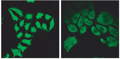
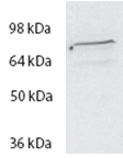
Application
| WB, IF, E |
|---|---|
| Primary Accession | Q8N0X7 |
| Other Accession | NP_055902.1, 23111 |
| Reactivity | Human |
| Host | Goat |
| Clonality | Polyclonal |
| Concentration | 0.5 mg/ml |
| Isotype | IgG |
| Calculated MW | 72833 Da |
| Gene ID | 23111 |
|---|---|
| Other Names | Spartin, Spastic paraplegia 20 protein, Trans-activated by hepatitis C virus core protein 1, SPG20, KIAA0610, TAHCCP1 |
| Dilution | WB~~1:1000 IF~~1:50~200 E~~N/A |
| Format | 0.5 mg/ml in Tris saline, 0.02% sodium azide, pH7.3 with 0.5% bovine serum albumin |
| Storage | Maintain refrigerated at 2-8°C for up to 6 months. For long term storage store at -20°C in small aliquots to prevent freeze-thaw cycles. |
| Precautions | SPARTIN Antibody (internal region) is for research use only and not for use in diagnostic or therapeutic procedures. |
| Name | SPART (HGNC:18514) |
|---|---|
| Function | Lipophagy receptor that plays an important role in lipid droplet (LD) turnover in motor neurons (PubMed:37443287). Localizes to LDs and interacts with components of the autophagy machinery, such as MAP1LC3A/C proteins to deliver LDs to autophagosomes for degradation via lipophagy (PubMed:37443287). Lipid transfer protein required for lipid droplet degradation, including by lipophagy (PubMed:38190532). Can bind and transfer all lipid species found in lipid droplets, from phospholipids to triglycerides and sterol esters but the direction of lipid transfer by spartin and its cargos are unknown (PubMed:38190532). May be implicated in endosomal trafficking, or microtubule dynamics, or both. Participates in cytokinesis (PubMed:20719964). |
| Cellular Location | Cytoplasm. Midbody. Lipid droplet Note=Transiently associated with endosomes (PubMed:19580544) Colocalized with IST1 to the ends of Flemming bodies during cytokinesis (PubMed:20719964). |
| Tissue Location | Ubiquitously expressed, with highest levels of expression detected in adipose tissue |

Thousands of laboratories across the world have published research that depended on the performance of antibodies from Abcepta to advance their research. Check out links to articles that cite our products in major peer-reviewed journals, organized by research category.
info@abcepta.com, and receive a free "I Love Antibodies" mug.

Provided below are standard protocols that you may find useful for product applications.
References
The hereditary spastic paraplegia protein spartin localises to mitochondria Lu J, Rashid F, Byrne PC. J Neurochem. 2006 Sep;98(6):1908-19. PMID: 16945107
If you have used an Abcepta product and would like to share how it has performed, please click on the "Submit Review" button and provide the requested information. Our staff will examine and post your review and contact you if needed.
If you have any additional inquiries please email technical services at tech@abcepta.com.
Ordering Information
Other Products
Shipping Information
