


 Foundational characteristics of cancer include proliferation, angiogenesis, migration, evasion of apoptosis, and cellular immortality. Find key markers for these cellular processes and antibodies to detect them.
 The SUMOplot™ Analysis Program predicts and scores sumoylation sites in your protein. SUMOylation is a post-translational modification involved in various cellular processes, such as nuclear-cytosolic transport, transcriptional regulation, apoptosis, protein stability, response to stress, and progression through the cell cycle.
 The Autophagy Receptor Motif Plotter predicts and scores autophagy receptor binding sites in your protein. Identifying proteins connected to this pathway is critical to understanding the role of autophagy in physiological as well as pathological processes such as development, differentiation, neurodegenerative diseases, stress, infection, and cancer.
EDG3 Antibody (internal region)
Peptide-affinity purified goat antibody
- SPECIFICATION
- CITATIONS
- PROTOCOLS
- BACKGROUND

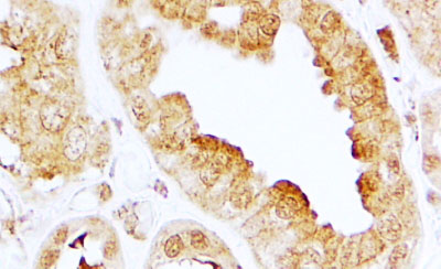
Application
| IHC, E |
|---|---|
| Primary Accession | Q99500 |
| Other Accession | NP_005217.2, 1903 |
| Reactivity | Human |
| Host | Goat |
| Clonality | Polyclonal |
| Concentration | 0.5 mg/ml |
| Isotype | IgG |
| Calculated MW | 42250 Da |
| Gene ID | 1903 |
|---|---|
| Other Names | Sphingosine 1-phosphate receptor 3, S1P receptor 3, S1P3, Endothelial differentiation G-protein coupled receptor 3, Sphingosine 1-phosphate receptor Edg-3, S1P receptor Edg-3, S1PR3, EDG3 |
| Dilution | IHC~~1:100~500 E~~N/A |
| Format | 0.5 mg/ml in Tris saline, 0.02% sodium azide, pH7.3 with 0.5% bovine serum albumin |
| Storage | Maintain refrigerated at 2-8°C for up to 6 months. For long term storage store at -20°C in small aliquots to prevent freeze-thaw cycles. |
| Precautions | EDG3 Antibody (internal region) is for research use only and not for use in diagnostic or therapeutic procedures. |
| Name | S1PR3 (HGNC:3167) |
|---|---|
| Function | Receptor for the lysosphingolipid sphingosine 1-phosphate (S1P). S1P is a bioactive lysophospholipid that elicits diverse physiological effect on most types of cells and tissues. When expressed in rat HTC4 hepatoma cells, is capable of mediating S1P-induced cell proliferation and suppression of apoptosis. |
| Cellular Location | Cell membrane; Multi-pass membrane protein. |
| Tissue Location | Expressed in all tissues, but most abundantly in heart, placenta, kidney, and liver |

Thousands of laboratories across the world have published research that depended on the performance of antibodies from Abcepta to advance their research. Check out links to articles that cite our products in major peer-reviewed journals, organized by research category.
info@abcepta.com, and receive a free "I Love Antibodies" mug.

Provided below are standard protocols that you may find useful for product applications.
References
Dendritic cell PAR1-S1P3 signalling couples coagulation and inflammation Niessen F, Schaffner F, Furlan-Freguia C, Pawlinski R, Bhattacharjee G, Chun J, Derian CK, Andrade-Gordon P, Rosen H, Ruf W Nature. 2008 Apr 3;452(7187):654-8. Epub 2008 Feb 27 PMID: 18305483
If you have used an Abcepta product and would like to share how it has performed, please click on the "Submit Review" button and provide the requested information. Our staff will examine and post your review and contact you if needed.
If you have any additional inquiries please email technical services at tech@abcepta.com.
Ordering Information
Other Products
Shipping Information
