


 Foundational characteristics of cancer include proliferation, angiogenesis, migration, evasion of apoptosis, and cellular immortality. Find key markers for these cellular processes and antibodies to detect them.
 The SUMOplot™ Analysis Program predicts and scores sumoylation sites in your protein. SUMOylation is a post-translational modification involved in various cellular processes, such as nuclear-cytosolic transport, transcriptional regulation, apoptosis, protein stability, response to stress, and progression through the cell cycle.
 The Autophagy Receptor Motif Plotter predicts and scores autophagy receptor binding sites in your protein. Identifying proteins connected to this pathway is critical to understanding the role of autophagy in physiological as well as pathological processes such as development, differentiation, neurodegenerative diseases, stress, infection, and cancer.
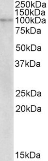
NLRX1 / NOD9 Antibody (internal region)
Peptide-affinity purified goat antibody
- SPECIFICATION
- CITATIONS
- PROTOCOLS
- BACKGROUND

Application
| WB, E |
|---|---|
| Primary Accession | Q86UT6 |
| Other Accession | NP_078894.2, NP_733840.1, 79671 |
| Reactivity | Human |
| Predicted | Rat |
| Host | Goat |
| Clonality | Polyclonal |
| Concentration | 0.5 mg/ml |
| Isotype | IgG |
| Calculated MW | 107616 Da |
| Gene ID | 79671 |
|---|---|
| Other Names | NLR family member X1, Caterpiller protein 11.3, CLR11.3, Nucleotide-binding oligomerization domain protein 26, Nucleotide-binding oligomerization domain protein 5, Nucleotide-binding oligomerization domain protein 9, NLRX1, NOD26, NOD5, NOD9 |
| Dilution | WB~~1:1000 E~~N/A |
| Format | 0.5 mg/ml in Tris saline, 0.02% sodium azide, pH7.3 with 0.5% bovine serum albumin |
| Storage | Maintain refrigerated at 2-8°C for up to 6 months. For long term storage store at -20°C in small aliquots to prevent freeze-thaw cycles. |
| Precautions | NLRX1 / NOD9 Antibody (internal region) is for research use only and not for use in diagnostic or therapeutic procedures. |
| Name | NLRX1 |
|---|---|
| Function | Participates in antiviral signaling. Acts as a negative regulator of MAVS-mediated antiviral responses, through the inhibition of the virus-induced RLH (RIG-like helicase)-MAVS interaction (PubMed:18200010). Instead, promotes autophagy by interacting with TUFM and subsequently recruiting the autophagy-related proteins ATG5 and ATG12 (PubMed:22749352). Also regulates MAVS-dependent NLRP3 inflammasome activation to attenuate apoptosis (PubMed:27393910). Has no inhibitory function on NF-kappa-B signaling pathway, but enhances NF-kappa-B and JUN N-terminal kinase dependent signaling through the production of reactive oxygen species (PubMed:18219313). Regulates viral mediated-inflammation and energy metabolism in a sex-dependent manner (By similarity). In females, prevents uncontrolled inflammation and energy metabolism and thus, may contribute to the sex differences observed in infectious and inflammatory diseases (By similarity). |
| Cellular Location | Mitochondrion outer membrane |
| Tissue Location | Ubiquitously expressed. Strongest expression in mammary gland, heart and muscle. Detected in HeLa, HEK293T, THP-1, HL- 60, Raji and Jurkat cell lines (at protein level) |

Thousands of laboratories across the world have published research that depended on the performance of antibodies from Abcepta to advance their research. Check out links to articles that cite our products in major peer-reviewed journals, organized by research category.
info@abcepta.com, and receive a free "I Love Antibodies" mug.

Provided below are standard protocols that you may find useful for product applications.
Background
This antibody is expected to recognize both reported isoforms (NP_078894.2; NP_733840.1).
References
NLRX1 is a mitochondrial NOD-like receptor that amplifies NF-kappaB and JNK pathways by inducing reactive oxygen species production. Tattoli I, Carneiro LA, Jéhanno M, Magalhaes JG, Shu Y, Philpott DJ, Arnoult D, Girardin SE, EMBO reports 2008 Mar 9 (3): 293-300. PMID: 18219313
If you have used an Abcepta product and would like to share how it has performed, please click on the "Submit Review" button and provide the requested information. Our staff will examine and post your review and contact you if needed.
If you have any additional inquiries please email technical services at tech@abcepta.com.
Ordering Information
Other Products
Shipping Information
