


 Foundational characteristics of cancer include proliferation, angiogenesis, migration, evasion of apoptosis, and cellular immortality. Find key markers for these cellular processes and antibodies to detect them.
 The SUMOplot™ Analysis Program predicts and scores sumoylation sites in your protein. SUMOylation is a post-translational modification involved in various cellular processes, such as nuclear-cytosolic transport, transcriptional regulation, apoptosis, protein stability, response to stress, and progression through the cell cycle.
 The Autophagy Receptor Motif Plotter predicts and scores autophagy receptor binding sites in your protein. Identifying proteins connected to this pathway is critical to understanding the role of autophagy in physiological as well as pathological processes such as development, differentiation, neurodegenerative diseases, stress, infection, and cancer.
OXER1 (aa389-400) Antibody (C-Term)
Peptide-affinity purified goat antibody
- SPECIFICATION
- CITATIONS
- PROTOCOLS
- BACKGROUND

Application
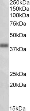
| WB, E |
|---|---|
| Primary Accession | Q8TDS5 |
| Other Accession | NP_683765.1, 165140 |
| Reactivity | Human |
| Host | Goat |
| Clonality | Polyclonal |
| Concentration | 0.5 mg/ml |
| Isotype | IgG |
| Calculated MW | 41426 Da |
| Gene ID | 165140 |
|---|---|
| Other Names | Oxoeicosanoid receptor 1, 5-oxo-ETE G-protein coupled receptor, G-protein coupled receptor 170, G-protein coupled receptor R527, G-protein coupled receptor TG1019, OXER1, GPR170, TG1019 |
| Dilution | WB~~1:1000 E~~N/A |
| Format | 0.5 mg/ml in Tris saline, 0.02% sodium azide, pH7.3 with 0.5% bovine serum albumin |
| Storage | Maintain refrigerated at 2-8°C for up to 6 months. For long term storage store at -20°C in small aliquots to prevent freeze-thaw cycles. |
| Precautions | OXER1 (aa389-400) Antibody (C-Term) is for research use only and not for use in diagnostic or therapeutic procedures. |
| Name | OXER1 |
|---|---|
| Synonyms | GPR170, TG1019 |
| Function | Receptor for eicosanoids and polyunsaturated fatty acids such as 5-oxo-6E,8Z,11Z,14Z-eicosatetraenoic acid (5-OXO-ETE), 5(S)- hydroperoxy-6E,8Z,11Z,14Z-eicosatetraenoic acid (5(S)-HPETE) and arachidonic acid. Seems to be coupled to the G(i)/G(o), families of heteromeric G proteins. |
| Cellular Location | Membrane; Multi-pass membrane protein |
| Tissue Location | Expressed in various tissues except brain. Expression is more intense in liver, kidney, peripheral leukocyte, lung, and spleen than in other tissues. Highly expressed in eosinophils, neutrophils, and lung macrophages |

Thousands of laboratories across the world have published research that depended on the performance of antibodies from Abcepta to advance their research. Check out links to articles that cite our products in major peer-reviewed journals, organized by research category.
info@abcepta.com, and receive a free "I Love Antibodies" mug.

Provided below are standard protocols that you may find useful for product applications.
References
Identification of a novel human eicosanoid receptor coupled to G(i/o). Hosoi T, Koguchi Y, Sugikawa E, Chikada A, Ogawa K, Tsuda N, Suto N, Tsunoda S, Taniguchi T, Ohnuki T. J Biol Chem. 2002 Aug 30;277(35):31459-65. PMID: 12065583
If you have used an Abcepta product and would like to share how it has performed, please click on the "Submit Review" button and provide the requested information. Our staff will examine and post your review and contact you if needed.
If you have any additional inquiries please email technical services at tech@abcepta.com.
Ordering Information
Other Products
Shipping Information
