


 Foundational characteristics of cancer include proliferation, angiogenesis, migration, evasion of apoptosis, and cellular immortality. Find key markers for these cellular processes and antibodies to detect them.
 The SUMOplot™ Analysis Program predicts and scores sumoylation sites in your protein. SUMOylation is a post-translational modification involved in various cellular processes, such as nuclear-cytosolic transport, transcriptional regulation, apoptosis, protein stability, response to stress, and progression through the cell cycle.
 The Autophagy Receptor Motif Plotter predicts and scores autophagy receptor binding sites in your protein. Identifying proteins connected to this pathway is critical to understanding the role of autophagy in physiological as well as pathological processes such as development, differentiation, neurodegenerative diseases, stress, infection, and cancer.
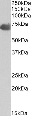
C9 (aa205-216) Antibody (internal region)
Peptide-affinity purified goat antibody
- SPECIFICATION
- CITATIONS
- PROTOCOLS
- BACKGROUND

Application
| WB, E |
|---|---|
| Primary Accession | P02748 |
| Other Accession | NP_001728.1, 735 |
| Reactivity | Human |
| Host | Goat |
| Clonality | Polyclonal |
| Concentration | 0.5 mg/ml |
| Isotype | IgG |
| Calculated MW | 63173 Da |
| Gene ID | 735 |
|---|---|
| Other Names | Complement component C9, Complement component C9a, Complement component C9b, C9 |
| Dilution | WB~~1:1000 E~~N/A |
| Format | 0.5 mg/ml in Tris saline, 0.02% sodium azide, pH7.3 with 0.5% bovine serum albumin |
| Storage | Maintain refrigerated at 2-8°C for up to 6 months. For long term storage store at -20°C in small aliquots to prevent freeze-thaw cycles. |
| Precautions | C9 (aa205-216) Antibody (internal region) is for research use only and not for use in diagnostic or therapeutic procedures. |
| Name | C9 {ECO:0000303|PubMed:4018030, ECO:0000312|HGNC:HGNC:1358} |
|---|---|
| Function | Pore-forming component of the membrane attack complex (MAC), a multiprotein complex activated by the complement cascade, which inserts into a target cell membrane and forms a pore, leading to target cell membrane rupture and cell lysis (PubMed:22832194, PubMed:26841837, PubMed:26841934, PubMed:27052168, PubMed:30552328, PubMed:6177822, PubMed:9212048, PubMed:9634479). The MAC is initiated by proteolytic cleavage of C5 into complement C5b in response to the classical, alternative, lectin and GZMK complement pathways (PubMed:9212048, PubMed:9634479). The complement pathways consist in a cascade of proteins that leads to phagocytosis and breakdown of pathogens and signaling that strengthens the adaptive immune system (PubMed:9212048, PubMed:9634479). Constitutes the pore-forming subunit of the MAC complex: during MAC assembly, C9 associates with the C5b8 intermediate complex, and polymerizes to complete the pore (PubMed:26841934, PubMed:30111885, PubMed:30552328, PubMed:34752492, PubMed:4055801, PubMed:6177822). |
| Cellular Location | Secreted. Target cell membrane; Multi-pass membrane protein. Note=Secreted as soluble monomer (PubMed:26841934, PubMed:30111885, PubMed:4055801, PubMed:9634479) Oligomerizes at target membranes, forming a pre-pore (PubMed:26841934, PubMed:30111885, PubMed:31061395, PubMed:4055801, PubMed:9634479). A conformation change then leads to the formation of a 100 Angstrom diameter pore (PubMed:26841934, PubMed:30111885, PubMed:31061395, PubMed:4055801, PubMed:9634479). |
| Tissue Location | Plasma (at protein level). |

Thousands of laboratories across the world have published research that depended on the performance of antibodies from Abcepta to advance their research. Check out links to articles that cite our products in major peer-reviewed journals, organized by research category.
info@abcepta.com, and receive a free "I Love Antibodies" mug.

Provided below are standard protocols that you may find useful for product applications.
References
Mapping the intermedilysin-human CD59 receptor interface reveals a deep correspondence with the binding site on CD59 for complement binding proteins C8alpha and C9. Wickham SE, Hotze EM, Farrand AJ, Polekhina G, Nero TL, Tomlinson S, Parker MW, Tweten RK. The Journal of biological chemistry 2011 Jun 286 (23): 20952-62. PMID: 21507937
If you have used an Abcepta product and would like to share how it has performed, please click on the "Submit Review" button and provide the requested information. Our staff will examine and post your review and contact you if needed.
If you have any additional inquiries please email technical services at tech@abcepta.com.
Ordering Information
Other Products
Shipping Information
