


 Foundational characteristics of cancer include proliferation, angiogenesis, migration, evasion of apoptosis, and cellular immortality. Find key markers for these cellular processes and antibodies to detect them.
 The SUMOplot™ Analysis Program predicts and scores sumoylation sites in your protein. SUMOylation is a post-translational modification involved in various cellular processes, such as nuclear-cytosolic transport, transcriptional regulation, apoptosis, protein stability, response to stress, and progression through the cell cycle.
 The Autophagy Receptor Motif Plotter predicts and scores autophagy receptor binding sites in your protein. Identifying proteins connected to this pathway is critical to understanding the role of autophagy in physiological as well as pathological processes such as development, differentiation, neurodegenerative diseases, stress, infection, and cancer.
OAS1 Antibody (internal region, near C-Term)
Peptide-affinity purified goat antibody
- SPECIFICATION
- CITATIONS
- PROTOCOLS
- BACKGROUND

Application
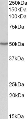
| WB, E |
|---|---|
| Primary Accession | P00973 |
| Other Accession | NP_058132.2, 4938 |
| Reactivity | Human |
| Host | Goat |
| Clonality | Polyclonal |
| Concentration | 0.5 mg/ml |
| Isotype | IgG |
| Calculated MW | 46029 Da |
| Gene ID | 4938 |
|---|---|
| Other Names | 2'-5'-oligoadenylate synthase 1, (2-5')oligo(A) synthase 1, 2-5A synthase 1, 2.7.7.84, E18/E16, p46/p42 OAS, OAS1, OIAS |
| Dilution | WB~~1:1000 E~~N/A |
| Format | 0.5 mg/ml in Tris saline, 0.02% sodium azide, pH7.3 with 0.5% bovine serum albumin |
| Storage | Maintain refrigerated at 2-8°C for up to 6 months. For long term storage store at -20°C in small aliquots to prevent freeze-thaw cycles. |
| Precautions | OAS1 Antibody (internal region, near C-Term) is for research use only and not for use in diagnostic or therapeutic procedures. |
| Name | OAS1 |
|---|---|
| Synonyms | OIAS |
| Function | Interferon-induced, dsRNA-activated antiviral enzyme which plays a critical role in cellular innate antiviral response (PubMed:34581622). In addition, it may also play a role in other cellular processes such as apoptosis, cell growth, differentiation and gene regulation. Synthesizes higher oligomers of 2'-5'-oligoadenylates (2-5A) from ATP which then bind to the inactive monomeric form of ribonuclease L (RNase L) leading to its dimerization and subsequent activation. Activation of RNase L leads to degradation of cellular as well as viral RNA, resulting in the inhibition of protein synthesis, thus terminating viral replication (PubMed:34145065, PubMed:34581622). Can mediate the antiviral effect via the classical RNase L-dependent pathway or an alternative antiviral pathway independent of RNase L. The secreted form displays antiviral effect against vesicular stomatitis virus (VSV), herpes simplex virus type 2 (HSV-2), and encephalomyocarditis virus (EMCV) and stimulates the alternative antiviral pathway independent of RNase L. |
| Cellular Location | Cytoplasm. Mitochondrion. Nucleus. Microsome Endoplasmic reticulum. Secreted {ECO:0000250|UniProtKB:Q29599}. Note=Associated with different subcellular fractions such as mitochondrial, nuclear, and rough/smooth microsomal fractions. [Isoform p42]: Note=(Microbial infection) In SARS coronavirus-2/SARS-CoV-2 infected cells, since its not prenylated, is diffusely localized and unable to initiate a detectable block to SARS- CoV-2 replication. |
| Tissue Location | Expressed in lungs.. |

Thousands of laboratories across the world have published research that depended on the performance of antibodies from Abcepta to advance their research. Check out links to articles that cite our products in major peer-reviewed journals, organized by research category.
info@abcepta.com, and receive a free "I Love Antibodies" mug.

Provided below are standard protocols that you may find useful for product applications.
Background
This antibody is expected to recognize isoforms 1 (NP_058132.2) only.
References
Polymorphism of OAS-1 determines liver fibrosis progression in hepatitis C by reduced ability to inhibit viral replication. Li CZ, Kato N, Chang JH, Muroyama R, Shao RX, Dharel N, Sermsathanasawadi R, Kawabe T, Omata M. Liver international : official journal of the International Association for the Study of the Liver 2009 Oct 29 (9): 1413-21. PMID: 19515215
If you have used an Abcepta product and would like to share how it has performed, please click on the "Submit Review" button and provide the requested information. Our staff will examine and post your review and contact you if needed.
If you have any additional inquiries please email technical services at tech@abcepta.com.
Ordering Information
Other Products
Shipping Information
