


 Foundational characteristics of cancer include proliferation, angiogenesis, migration, evasion of apoptosis, and cellular immortality. Find key markers for these cellular processes and antibodies to detect them.
 The SUMOplot™ Analysis Program predicts and scores sumoylation sites in your protein. SUMOylation is a post-translational modification involved in various cellular processes, such as nuclear-cytosolic transport, transcriptional regulation, apoptosis, protein stability, response to stress, and progression through the cell cycle.
 The Autophagy Receptor Motif Plotter predicts and scores autophagy receptor binding sites in your protein. Identifying proteins connected to this pathway is critical to understanding the role of autophagy in physiological as well as pathological processes such as development, differentiation, neurodegenerative diseases, stress, infection, and cancer.
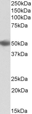
Goat Anti-DC-SIGN / CD209 Antibody (internal region)
Purified Goat Polyclonal Antibody
- SPECIFICATION
- CITATIONS
- PROTOCOLS
- BACKGROUND

Application
| WB, E |
|---|---|
| Primary Accession | Q9NNX6 |
| Other Accession | NP_066978.1, NP_001138368.1, NP_001138369.1, NP_001138365.1, NP_001138366.1, NP_001138367.1, NP_001138371.1 |
| Reactivity | Human |
| Predicted | Human |
| Host | Goat |
| Clonality | Polyclonal |
| Concentration | 0.5 |
| Calculated MW | 45775 Da |
| Gene ID | 30835 |
|---|---|
| Other Names | CD209; CD209 molecule; CDSIGN; CLEC4L; DC-SIGN; DC-SIGN1; C-type lectin domain family 4 member L; C-type lectin domain family 4, member L; CD209 antigen; HIV gpl20-binding protein; dendritic cell-specific ICAM-3-grabbing non-integrin 1; dendritic cell-spe |
| Dilution | WB~~1:1000 E~~N/A |
| Format | Supplied at 0.5 mg/ml in Tris saline, 0.02% sodium azide, pH7.3 with 0.5% bovine serum albumin. Aliquot and store at -20°C. Minimize freezing and thawing. |
| Immunogen | Peptide with sequence C-NRGEPNNVGEED, from the internal region of the protein sequence according to NP_066978.1; NP_001138368.1; NP_001138369.1; NP_001138365.1; NP_001138366.1; NP_001138367.1; NP_001138371.1. |
| Storage | Maintain refrigerated at 2-8°C for up to 6 months. For long term storage store at -20°C in small aliquots to prevent freeze-thaw cycles. |
| Precautions | Goat Anti-DC-SIGN / CD209 Antibody (internal region) is for research use only and not for use in diagnostic or therapeutic procedures. |
| Name | CD209 |
|---|---|
| Synonyms | CLEC4L |
| Function | Pathogen-recognition receptor expressed on the surface of immature dendritic cells (DCs) and involved in initiation of primary immune response. Thought to mediate the endocytosis of pathogens which are subsequently degraded in lysosomal compartments. The receptor returns to the cell membrane surface and the pathogen-derived antigens are presented to resting T-cells via MHC class II proteins to initiate the adaptive immune response. |
| Cellular Location | [Isoform 1]: Cell membrane; Single- pass type II membrane protein [Isoform 3]: Cell membrane; Single- pass type II membrane protein [Isoform 5]: Cell membrane; Single- pass type II membrane protein [Isoform 7]: Secreted. [Isoform 9]: Secreted. [Isoform 11]: Secreted. |
| Tissue Location | Predominantly expressed in dendritic cells and in DC-residing tissues. Also found in placental macrophages, endothelial cells of placental vascular channels, peripheral blood mononuclear cells, and THP-1 monocytes. |

Thousands of laboratories across the world have published research that depended on the performance of antibodies from Abcepta to advance their research. Check out links to articles that cite our products in major peer-reviewed journals, organized by research category.
info@abcepta.com, and receive a free "I Love Antibodies" mug.

Provided below are standard protocols that you may find useful for product applications.
References
Role of DC-SIGN in Lassa virus entry into human dendritic cells. Goncalves AR, Moraz ML, Pasquato A, Helenius A, Lozach PY, Kunz S. Journal of virology 2013 Nov 87 (21): 11504-15.
If you have used an Abcepta product and would like to share how it has performed, please click on the "Submit Review" button and provide the requested information. Our staff will examine and post your review and contact you if needed.
If you have any additional inquiries please email technical services at tech@abcepta.com.
Ordering Information
Other Products
Shipping Information
