


 Foundational characteristics of cancer include proliferation, angiogenesis, migration, evasion of apoptosis, and cellular immortality. Find key markers for these cellular processes and antibodies to detect them.
 The SUMOplot™ Analysis Program predicts and scores sumoylation sites in your protein. SUMOylation is a post-translational modification involved in various cellular processes, such as nuclear-cytosolic transport, transcriptional regulation, apoptosis, protein stability, response to stress, and progression through the cell cycle.
 The Autophagy Receptor Motif Plotter predicts and scores autophagy receptor binding sites in your protein. Identifying proteins connected to this pathway is critical to understanding the role of autophagy in physiological as well as pathological processes such as development, differentiation, neurodegenerative diseases, stress, infection, and cancer.
> home > Products > Primary Antibodies > Antibody Collections > GPCR Antibodies > KD-Validated Anti-Frizzled class receptor 9 Rabbit Monoclonal Antibody
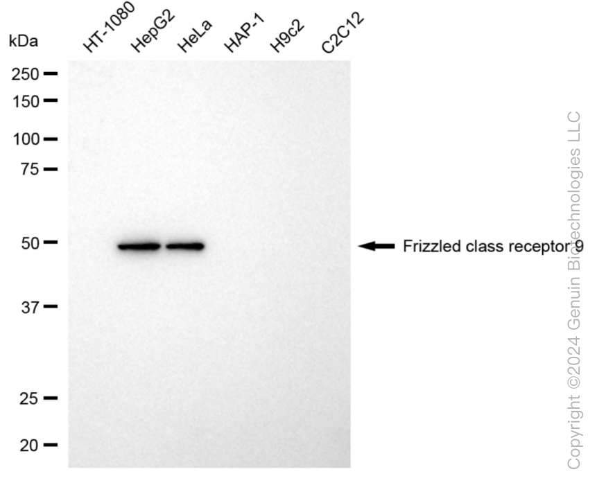
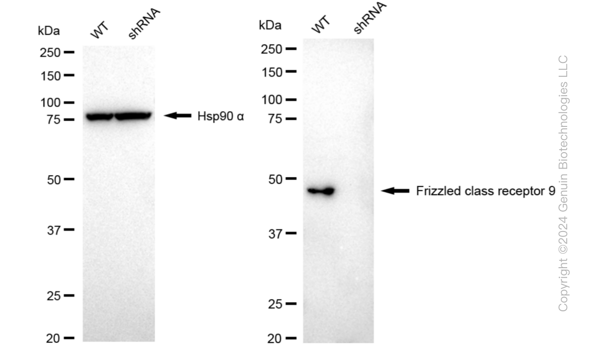
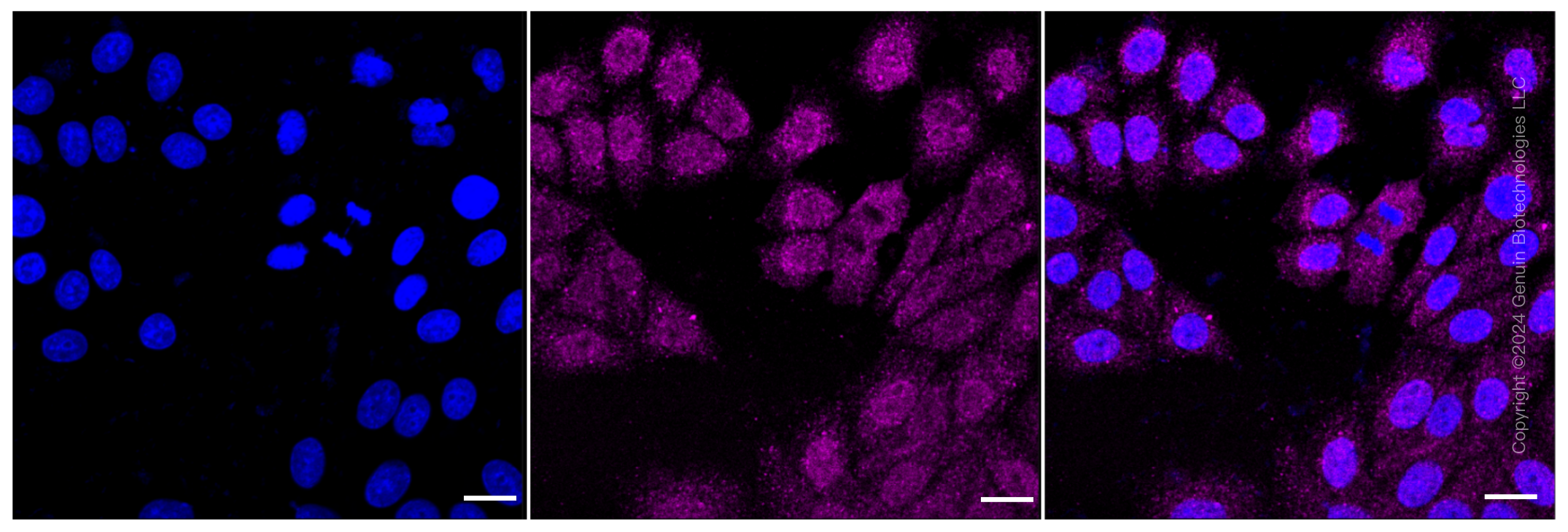
KD-Validated Anti-Frizzled class receptor 9 Rabbit Monoclonal Antibody
Rabbit monoclonal antibody
- SPECIFICATION
- CITATIONS
- PROTOCOLS
- BACKGROUND

Application
| WB, ICC |
|---|---|
| Primary Accession | O00144 |
| Reactivity | Human |
| Clonality | Monoclonal |
| Isotype | Rabbit IgG |
| Clone Names | 24GB5995 |
| Calculated MW | Predicted, 64 kDa, observed, 50 kDa |
| Gene Name | FZD9 |
| Aliases | FZD9; Frizzled Class Receptor 9; FZD3; CD349; Frizzled 9, Seven Transmembrane Spanning Receptor; Frizzled Family Receptor 9; Frizzled-9; Fz-9; FzE6; HFz9; Frizzled (Drosophila) Homolog 9; Frizzled Homolog 9 (Drosophila); Frizzled Homolog 9; CD349 Antigen |
| Immunogen | A synthesized peptide derived from human Frizzled 9 / CD349 |
| Gene ID | 8326 |
|---|---|
| Other Names | Frizzled-9, Fz-9, hFz9, FzE6, CD349, FZD9, FZD3 |
| Name | FZD9 |
|---|---|
| Synonyms | FZD3 |
| Function | Receptor for WNT2 that is coupled to the beta-catenin canonical signaling pathway, which leads to the activation of disheveled proteins, inhibition of GSK-3 kinase, nuclear accumulation of beta-catenin and activation of Wnt target genes (By similarity). Plays a role in neuromuscular junction (NMJ) assembly by negatively regulating the clustering of acetylcholine receptors (AChR) through the beta-catenin canonical signaling pathway (By similarity). May play a role in neural progenitor cells (NPCs) viability through the beta- catenin canonical signaling pathway by negatively regulating cell cycle arrest leading to inhibition of neuron apoptotic process (PubMed:27509850). During hippocampal development, regulates neuroblast proliferation and apoptotic cell death. Controls bone formation through non canonical Wnt signaling mediated via ISG15. Positively regulates bone regeneration through non canonical Wnt signaling (By similarity). |
| Cellular Location | Cell membrane {ECO:0000250|UniProtKB:Q9R216}; Multi-pass membrane protein. Note=Relocalizes DVL1 to the cell membrane leading to phosphorylation of DVL1 and AXIN1 relocalization to the cell membrane. {ECO:0000250|UniProtKB:Q8K4C8} |
| Tissue Location | Expressed predominantly in adult and fetal brain, testis, eye, skeletal muscle and kidney. Moderately expressed in pancreas, thyroid, adrenal cortex, small intestine and stomach Detected in fetal liver and kidney. Expressed in neural progenitor cells (PubMed:27509850). |
Research Areas
Citations (0)

Thousands of laboratories across the world have published research that depended on the performance of antibodies from Abcepta to advance their research. Check out links to articles that cite our products in major peer-reviewed journals, organized by research category.
Submit your citation using an Abcepta antibody to
info@abcepta.com, and receive a free "I Love Antibodies" mug.
info@abcepta.com, and receive a free "I Love Antibodies" mug.

Application Protocols
Provided below are standard protocols that you may find useful for product applications.
Abcepta welcomes feedback from its customers.
If you have used an Abcepta product and would like to share how it has performed, please click on the "Submit Review" button and provide the requested information. Our staff will examine and post your review and contact you if needed.
If you have any additional inquiries please email technical services at tech@abcepta.com.
$ 399.20

$ 149.00
Cat# AGI1034
Ordering Information
United States
AlbaniaAustraliaAustriaBelgiumBosnia & HerzegovinaBrazilBulgariaCanadaCentral AmericaChinaCroatiaCyprusCzech RepublicDenmarkEstoniaFinlandFranceGermanyGreeceHong KongHungaryIcelandIndiaIndonesiaIrelandIsraelItalyJapanLatviaLithuaniaLuxembourgMacedoniaMalaysiaMaltaMexicoNetherlandsNew ZealandNorwayPakistanPolandPortugalRomaniaSerbiaSingaporeSlovakiaSloveniaSouth AfricaSouth KoreaSpainSwedenSwitzerlandTaiwanTurkeyUnited KingdomUnited StatesVietnamWorldwideOthers
USA Headquarters
(888) 735-7227 / (858) 622-0099 or (858) 875-1900
Other Products
Shipping Information
Domestic orders (in stock items)
Shipped out the same day. Orders placed after 1 PM (PST) will ship out the next business day.
International orders
Contact your local distributors
