


 Foundational characteristics of cancer include proliferation, angiogenesis, migration, evasion of apoptosis, and cellular immortality. Find key markers for these cellular processes and antibodies to detect them.
 The SUMOplot™ Analysis Program predicts and scores sumoylation sites in your protein. SUMOylation is a post-translational modification involved in various cellular processes, such as nuclear-cytosolic transport, transcriptional regulation, apoptosis, protein stability, response to stress, and progression through the cell cycle.
 The Autophagy Receptor Motif Plotter predicts and scores autophagy receptor binding sites in your protein. Identifying proteins connected to this pathway is critical to understanding the role of autophagy in physiological as well as pathological processes such as development, differentiation, neurodegenerative diseases, stress, infection, and cancer.
> home > Products > Primary Antibodies > Antibody Collections > KD-Validated Antibodies > KD-Validated Anti-Cell division cycle 16 Rabbit Monoclonal Antibody
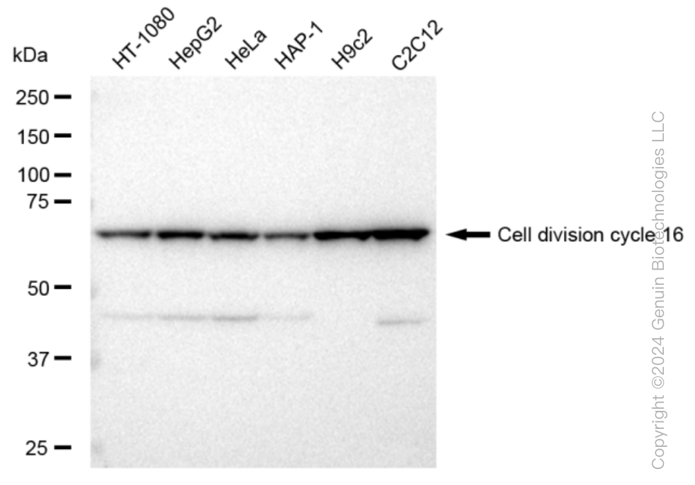
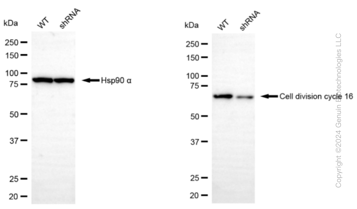
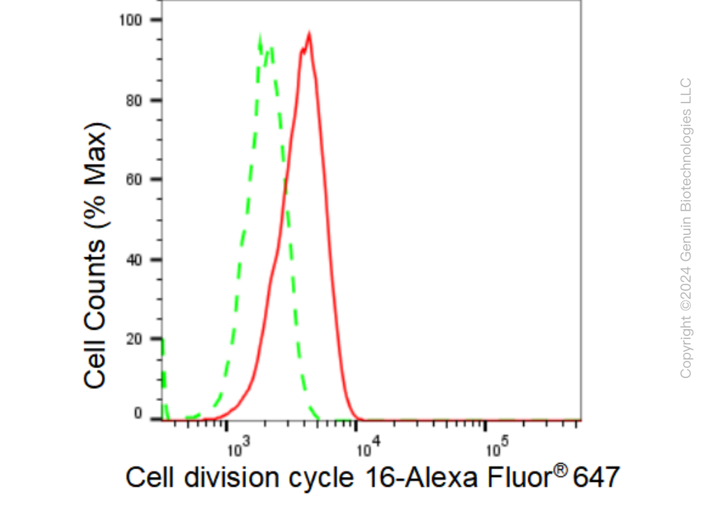
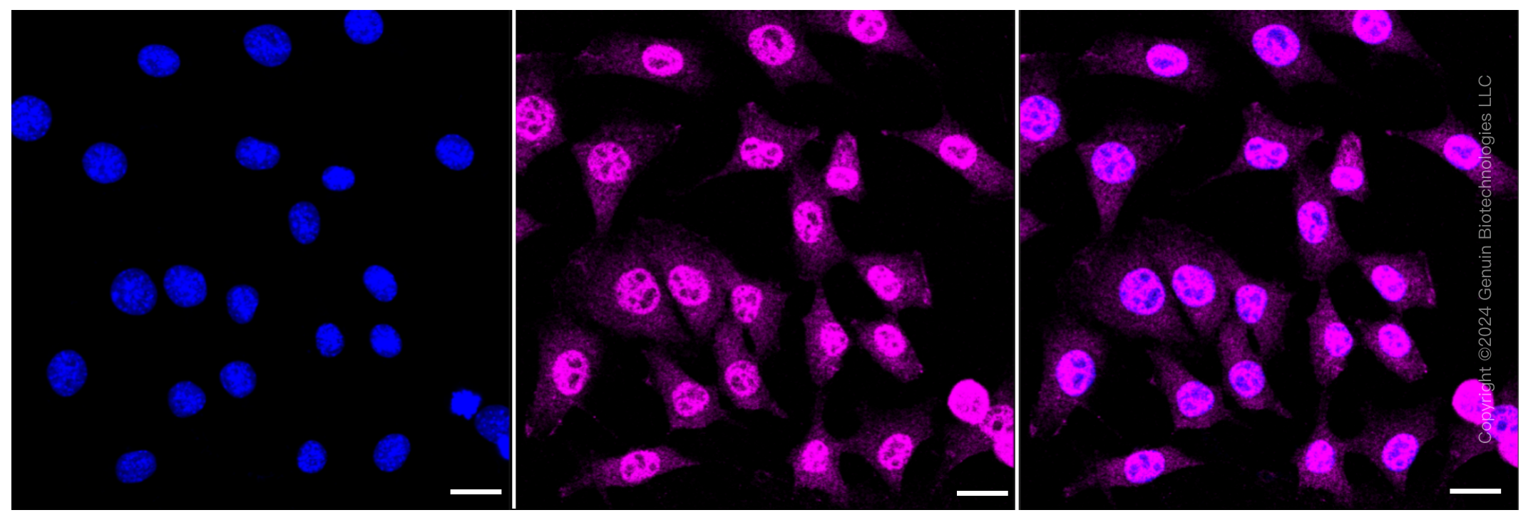
KD-Validated Anti-Cell division cycle 16 Rabbit Monoclonal Antibody
Rabbit monoclonal antibody
- SPECIFICATION
- CITATIONS
- PROTOCOLS
- BACKGROUND

Application
| WB, FC, ICC |
|---|---|
| Primary Accession | Q13042 |
| Reactivity | Rat, Human, Mouse |
| Clonality | Monoclonal |
| Isotype | Rabbit IgG |
| Clone Names | 23GB3455 |
| Calculated MW | Predicted, 72 kDa , observed, 71 kDa |
| Gene Name | CDC16 |
| Aliases | CDC16; Cell Division Cycle 16; ANAPC6; APC6; CUT9; Cell Division Cycle Protein 16 Homolog; Anaphase-Promoting Complex, Subunit 6; Cyclosome Subunit 6; CDC16Hs; CDC16 (Cell Division Cycle 16, S. Cerevisiae, Homolog); CDC16 Cell Division Cycle 16 Homolog (S. Cerevisiae); Cell Division Cycle 16 Homolog (S. Cerevisiae); Anaphase-Promoting Complex Subunit 6; Cell Division Cycle 16 Homolog; CDC16 Homolog |
| Immunogen | A synthesized peptide derived from human Apc6 / CDC16 |
| Gene ID | 8881 |
|---|---|
| Other Names | Cell division cycle protein 16 homolog, Anaphase-promoting complex subunit 6, APC6, CDC16 homolog, CDC16Hs, Cyclosome subunit 6, CDC16, ANAPC6 |
| Name | CDC16 |
|---|---|
| Synonyms | ANAPC6 |
| Function | Component of the anaphase promoting complex/cyclosome (APC/C), a cell cycle-regulated E3 ubiquitin ligase that controls progression through mitosis and the G1 phase of the cell cycle (PubMed:18485873). The APC/C complex acts by mediating ubiquitination and subsequent degradation of target proteins: it mainly mediates the formation of 'Lys-11'-linked polyubiquitin chains and, to a lower extent, the formation of 'Lys-48'- and 'Lys-63'-linked polyubiquitin chains (PubMed:18485873). The APC/C complex catalyzes assembly of branched 'Lys-11'-/'Lys-48'-linked branched ubiquitin chains on target proteins (PubMed:29033132). |
| Cellular Location | Cytoplasm, cytoskeleton, microtubule organizing center, centrosome. Cytoplasm, cytoskeleton, spindle. Note=Colocalizes with CDC27 to the centrosome at all stages of the cell cycle and to the mitotic spindle. |
Research Areas
Citations (0)

Thousands of laboratories across the world have published research that depended on the performance of antibodies from Abcepta to advance their research. Check out links to articles that cite our products in major peer-reviewed journals, organized by research category.
Submit your citation using an Abcepta antibody to
info@abcepta.com, and receive a free "I Love Antibodies" mug.
info@abcepta.com, and receive a free "I Love Antibodies" mug.

Application Protocols
Provided below are standard protocols that you may find useful for product applications.
Abcepta welcomes feedback from its customers.
If you have used an Abcepta product and would like to share how it has performed, please click on the "Submit Review" button and provide the requested information. Our staff will examine and post your review and contact you if needed.
If you have any additional inquiries please email technical services at tech@abcepta.com.
$ 149.00
$ 499.00
Cat# AGI1173
Ordering Information
United States
AlbaniaAustraliaAustriaBelgiumBosnia & HerzegovinaBrazilBulgariaCanadaCentral AmericaChinaCroatiaCyprusCzech RepublicDenmarkEstoniaFinlandFranceGermanyGreeceHong KongHungaryIcelandIndiaIndonesiaIrelandIsraelItalyJapanLatviaLithuaniaLuxembourgMacedoniaMalaysiaMaltaMexicoNetherlandsNew ZealandNorwayPakistanPolandPortugalRomaniaSerbiaSingaporeSlovakiaSloveniaSouth AfricaSouth KoreaSpainSwedenSwitzerlandTaiwanTurkeyUnited KingdomUnited StatesVietnamWorldwideOthers
USA Headquarters
(888) 735-7227 / (858) 622-0099 or (858) 875-1900
Other Products
Shipping Information
Domestic orders (in stock items)
Shipped out the same day. Orders placed after 1 PM (PST) will ship out the next business day.
International orders
Contact your local distributors
