


 Foundational characteristics of cancer include proliferation, angiogenesis, migration, evasion of apoptosis, and cellular immortality. Find key markers for these cellular processes and antibodies to detect them.
 The SUMOplot™ Analysis Program predicts and scores sumoylation sites in your protein. SUMOylation is a post-translational modification involved in various cellular processes, such as nuclear-cytosolic transport, transcriptional regulation, apoptosis, protein stability, response to stress, and progression through the cell cycle.
 The Autophagy Receptor Motif Plotter predicts and scores autophagy receptor binding sites in your protein. Identifying proteins connected to this pathway is critical to understanding the role of autophagy in physiological as well as pathological processes such as development, differentiation, neurodegenerative diseases, stress, infection, and cancer.
> home > Products > Primary Antibodies > Antibody Collections > KD-Validated Antibodies > KD-Validated Anti-WD repeat domain 1 Rabbit Monoclonal Antibody
KD-Validated Anti-WD repeat domain 1 Rabbit Monoclonal Antibody
Rabbit monoclonal antibody
- SPECIFICATION
- CITATIONS
- PROTOCOLS
- BACKGROUND

Application
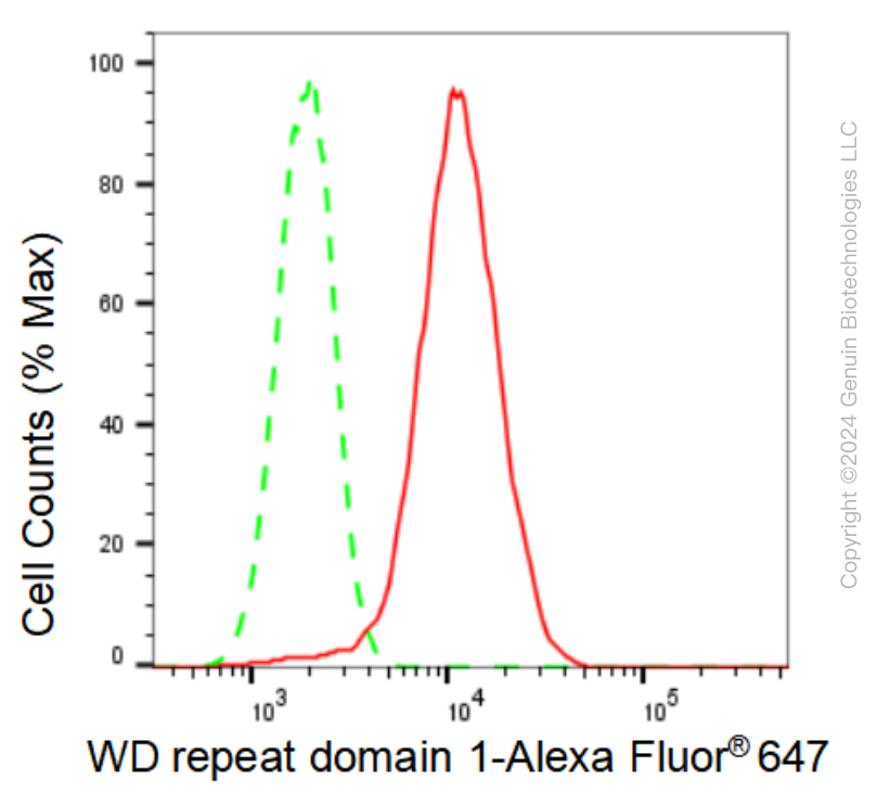
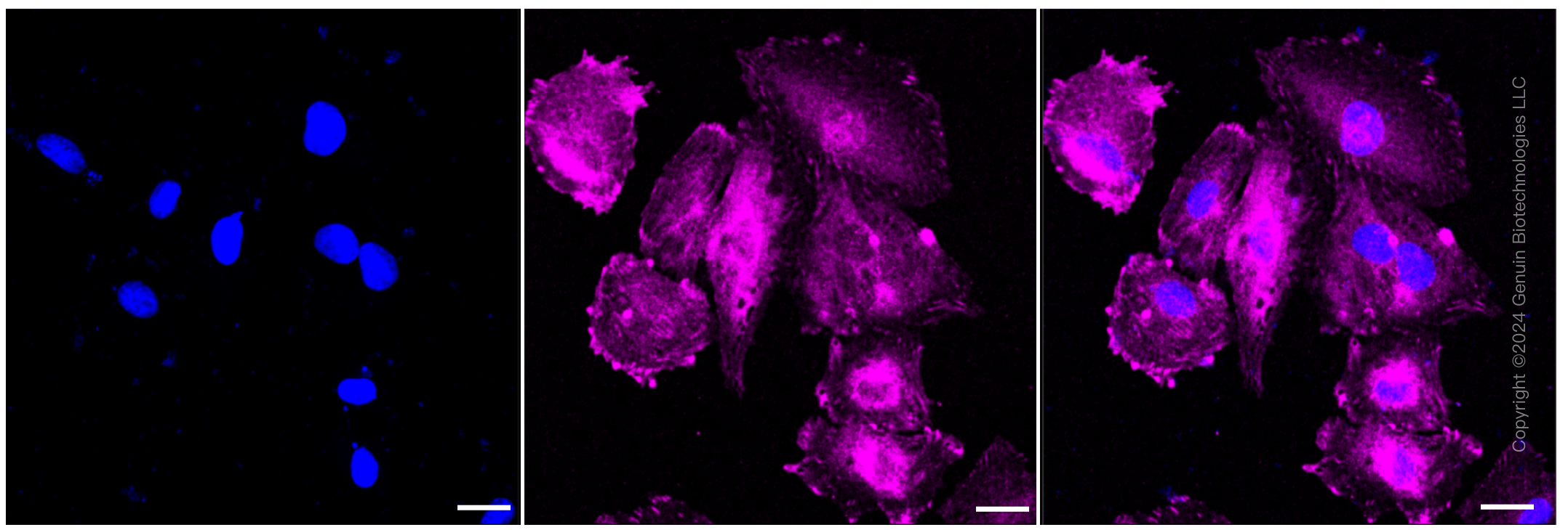
| WB, FC, ICC |
|---|---|
| Primary Accession | O75083 |
| Reactivity | Rat, Human, Mouse |
| Clonality | Monoclonal |
| Isotype | Rabbit IgG |
| Clone Names | 23GB4720 |
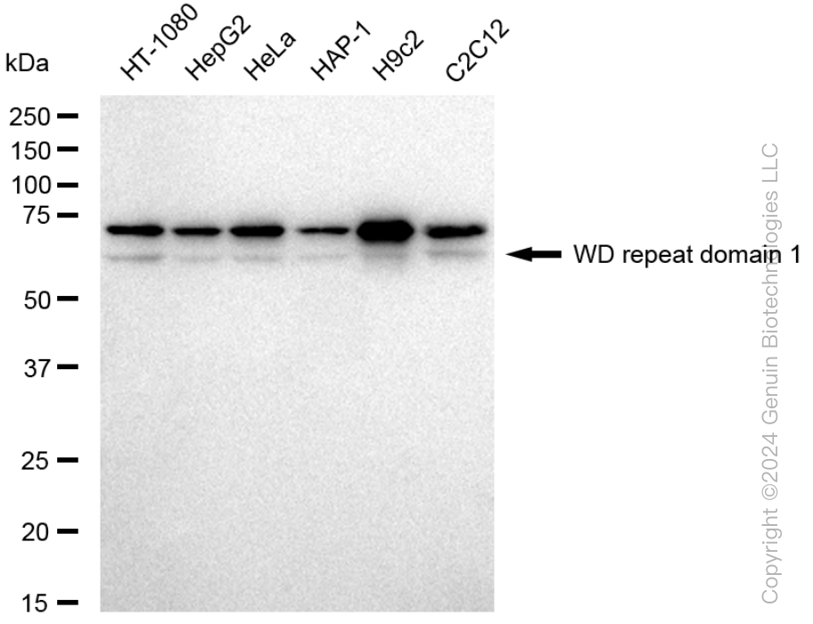
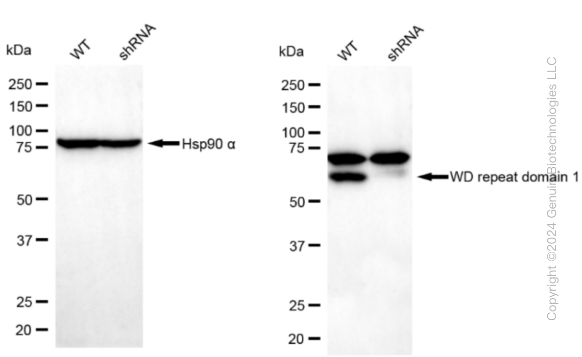
| Calculated MW | Predicted, 66 kDa, observed, 75 kDa |
| Gene Name | WDR1 |
| Aliases | WDR1; WD Repeat Domain 1; AIP1; Actin-Interacting Protein 1; WD Repeat-Containing Protein 1; NORI-1; Epididymis Secretory Protein Li 52; HEL-S-52; PFITS |
| Immunogen | A synthesized peptide derived from human WDR1 |
| Gene ID | 9948 |
|---|---|
| Other Names | WD repeat-containing protein 1, Actin-interacting protein 1, AIP1, NORI-1, WDR1 |
| Name | WDR1 |
|---|---|
| Function | Induces disassembly of actin filaments in conjunction with ADF/cofilin family proteins (PubMed:15629458, PubMed:27557945, PubMed:29751004). Enhances cofilin-mediated actin severing (By similarity). Involved in cytokinesis. Involved in chemotactic cell migration by restricting lamellipodial membrane protrusions (PubMed:18494608). Involved in myocardium sarcomere organization. Required for cardiomyocyte growth and maintenance (By similarity). Involved in megakaryocyte maturation and platelet shedding. Required for the establishment of planar cell polarity (PCP) during follicular epithelium development and for cell shape changes during PCP; the function seems to implicate cooperation with CFL1 and/or DSTN/ADF. Involved in the generation/maintenance of cortical tension (By similarity). Involved in assembly and maintenance of epithelial apical cell junctions and plays a role in the organization of the perijunctional actomyosin belt (PubMed:25792565). |
| Cellular Location | Cytoplasm. Cytoplasm, cytoskeleton {ECO:0000250|UniProtKB:Q5RKI0}. Cell projection, podosome. Cell junction |
| Tissue Location | Expressed in peripheral blood mononuclear cells (at protein level). |
Research Areas
Citations (0)

Thousands of laboratories across the world have published research that depended on the performance of antibodies from Abcepta to advance their research. Check out links to articles that cite our products in major peer-reviewed journals, organized by research category.
Submit your citation using an Abcepta antibody to
info@abcepta.com, and receive a free "I Love Antibodies" mug.
info@abcepta.com, and receive a free "I Love Antibodies" mug.

Application Protocols
Provided below are standard protocols that you may find useful for product applications.
Abcepta welcomes feedback from its customers.
If you have used an Abcepta product and would like to share how it has performed, please click on the "Submit Review" button and provide the requested information. Our staff will examine and post your review and contact you if needed.
If you have any additional inquiries please email technical services at tech@abcepta.com.
$ 399.20

$ 149.00
Cat# AGI1471
Ordering Information
United States
AlbaniaAustraliaAustriaBelgiumBosnia & HerzegovinaBrazilBulgariaCanadaCentral AmericaChinaCroatiaCyprusCzech RepublicDenmarkEstoniaFinlandFranceGermanyGreeceHong KongHungaryIcelandIndiaIndonesiaIrelandIsraelItalyJapanLatviaLithuaniaLuxembourgMacedoniaMalaysiaMaltaMexicoNetherlandsNew ZealandNorwayPakistanPolandPortugalRomaniaSerbiaSingaporeSlovakiaSloveniaSouth AfricaSouth KoreaSpainSwedenSwitzerlandTaiwanTurkeyUnited KingdomUnited StatesVietnamWorldwideOthers
USA Headquarters
(888) 735-7227 / (858) 622-0099 or (858) 875-1900
Other Products
Shipping Information
Domestic orders (in stock items)
Shipped out the same day. Orders placed after 1 PM (PST) will ship out the next business day.
International orders
Contact your local distributors
