


 Foundational characteristics of cancer include proliferation, angiogenesis, migration, evasion of apoptosis, and cellular immortality. Find key markers for these cellular processes and antibodies to detect them.
 The SUMOplot™ Analysis Program predicts and scores sumoylation sites in your protein. SUMOylation is a post-translational modification involved in various cellular processes, such as nuclear-cytosolic transport, transcriptional regulation, apoptosis, protein stability, response to stress, and progression through the cell cycle.
 The Autophagy Receptor Motif Plotter predicts and scores autophagy receptor binding sites in your protein. Identifying proteins connected to this pathway is critical to understanding the role of autophagy in physiological as well as pathological processes such as development, differentiation, neurodegenerative diseases, stress, infection, and cancer.
> home > Products > Primary Antibodies > Antibody Collections > KD-Validated Antibodies > KD-Validated Anti-Ubiquitination Factor E4B Rabbit Monoclonal Antibody
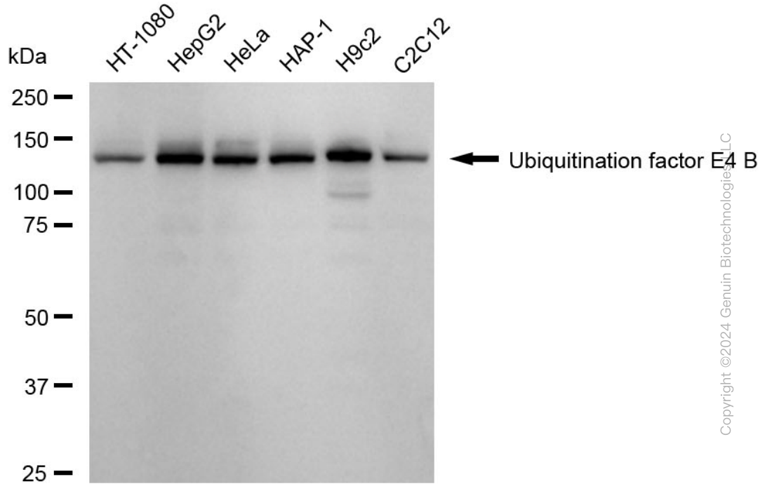
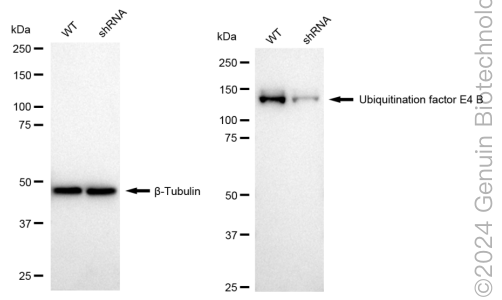
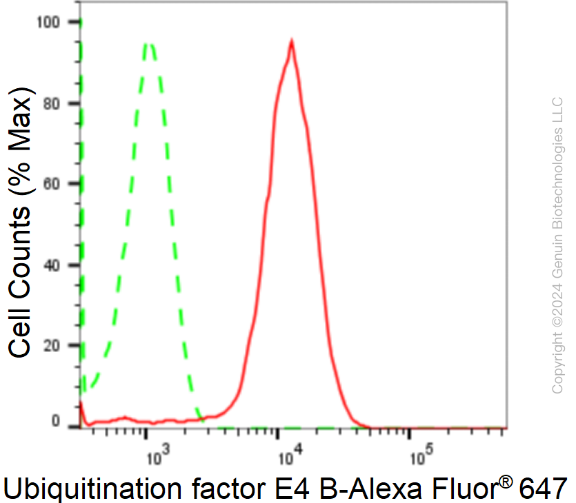
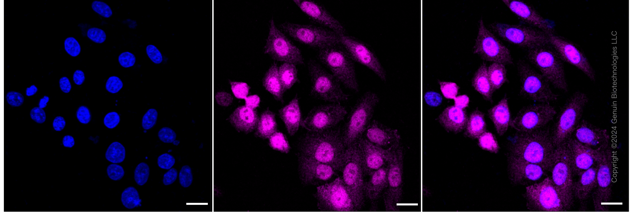
KD-Validated Anti-Ubiquitination Factor E4B Rabbit Monoclonal Antibody
Rabbit monoclonal antibody
- SPECIFICATION
- CITATIONS
- PROTOCOLS
- BACKGROUND

Application
| WB, FC, ICC |
|---|---|
| Primary Accession | O95155 |
| Reactivity | Rat, Human, Mouse |
| Clonality | Monoclonal |
| Isotype | Rabbit IgG |
| Clone Names | 24GB1765 |
| Calculated MW | Predicted, 146 kDa , observed , 130 kDa |
| Gene Name | UBE4B |
| Aliases | UBE4B; Ubiquitination Factor E4B; UFD2; KIAA0684; UBOX3; E4; Ubiquitination Factor E4B (UFD2 Homolog, Yeast); RING-Type E3 Ubiquitin Transferase E4 B; Ubiquitin Conjugation Factor E4 B; HDNB1; Ubiquitination Factor E4B (Homologous To Yeast UFD2); Homozygously Deleted In Neuroblastoma-1; Homozygously Deleted In Neuroblastoma 1; Ubiquitin-Fusion Degradation Protein 2; Ubiquitin Fusion Degradation Protein 2; Homologous To Yeast UFD2; EC 2.3.2.27; UFD2A |
| Immunogen | A synthesized peptide derived from human UBE4B |
| Gene ID | 10277 |
|---|---|
| Other Names | Ubiquitin conjugation factor E4 B, 2.3.2.27, UBE4B (HGNC:12500) |
| Name | UBE4B (HGNC:12500) |
|---|---|
| Function | Ubiquitin-protein ligase that probably functions as an E3 ligase in conjunction with specific E1 and E2 ligases (By similarity). May also function as an E4 ligase mediating the assembly of polyubiquitin chains on substrates ubiquitinated by another E3 ubiquitin ligase (By similarity). May regulate myosin assembly in striated muscles together with STUB1 and VCP/p97 by targeting myosin chaperone UNC45B for proteasomal degradation (PubMed:17369820). |
| Cellular Location | Cytoplasm. Nucleus {ECO:0000250|UniProtKB:Q9ES00} |
| Tissue Location | Expressed in differentiated myotubes (at protein level) (PubMed:17369820). Highest expression in ovary, testis, heart and skeletal muscle (PubMed:11802788). Expression is low in colon, thymus and peripheral blood leukocytes (PubMed:11802788). Almost undetectable in lung and spleen (PubMed:11802788) |
Research Areas
Citations (0)

Thousands of laboratories across the world have published research that depended on the performance of antibodies from Abcepta to advance their research. Check out links to articles that cite our products in major peer-reviewed journals, organized by research category.
Submit your citation using an Abcepta antibody to
info@abcepta.com, and receive a free "I Love Antibodies" mug.
info@abcepta.com, and receive a free "I Love Antibodies" mug.

Application Protocols
Provided below are standard protocols that you may find useful for product applications.
Abcepta welcomes feedback from its customers.
If you have used an Abcepta product and would like to share how it has performed, please click on the "Submit Review" button and provide the requested information. Our staff will examine and post your review and contact you if needed.
If you have any additional inquiries please email technical services at tech@abcepta.com.
$ 399.20

$ 149.00
Cat# AGI1716
Ordering Information
United States
AlbaniaAustraliaAustriaBelgiumBosnia & HerzegovinaBrazilBulgariaCanadaCentral AmericaChinaCroatiaCyprusCzech RepublicDenmarkEstoniaFinlandFranceGermanyGreeceHong KongHungaryIcelandIndiaIndonesiaIrelandIsraelItalyJapanLatviaLithuaniaLuxembourgMacedoniaMalaysiaMaltaMexicoNetherlandsNew ZealandNorwayPakistanPolandPortugalRomaniaSerbiaSingaporeSlovakiaSloveniaSouth AfricaSouth KoreaSpainSwedenSwitzerlandTaiwanTurkeyUnited KingdomUnited StatesVietnamWorldwideOthers
USA Headquarters
(888) 735-7227 / (858) 622-0099 or (858) 875-1900
Other Products
Shipping Information
Domestic orders (in stock items)
Shipped out the same day. Orders placed after 1 PM (PST) will ship out the next business day.
International orders
Contact your local distributors
