


 Foundational characteristics of cancer include proliferation, angiogenesis, migration, evasion of apoptosis, and cellular immortality. Find key markers for these cellular processes and antibodies to detect them.
 The SUMOplot™ Analysis Program predicts and scores sumoylation sites in your protein. SUMOylation is a post-translational modification involved in various cellular processes, such as nuclear-cytosolic transport, transcriptional regulation, apoptosis, protein stability, response to stress, and progression through the cell cycle.
 The Autophagy Receptor Motif Plotter predicts and scores autophagy receptor binding sites in your protein. Identifying proteins connected to this pathway is critical to understanding the role of autophagy in physiological as well as pathological processes such as development, differentiation, neurodegenerative diseases, stress, infection, and cancer.
> home > Products > Primary Antibodies > Antibody Collections > KD-Validated Antibodies > KD-Validated Anti-Epidermal Growth Factor Receptor Mouse Monoclonal Antibody
KD-Validated Anti-Epidermal Growth Factor Receptor Mouse Monoclonal Antibody
Mouse monoclonal antibody
- SPECIFICATION
- CITATIONS
- PROTOCOLS
- BACKGROUND

Application
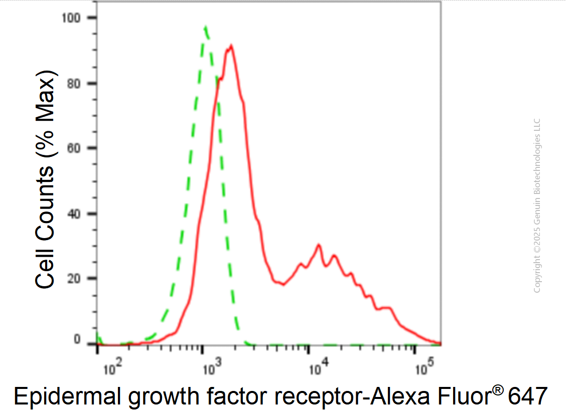
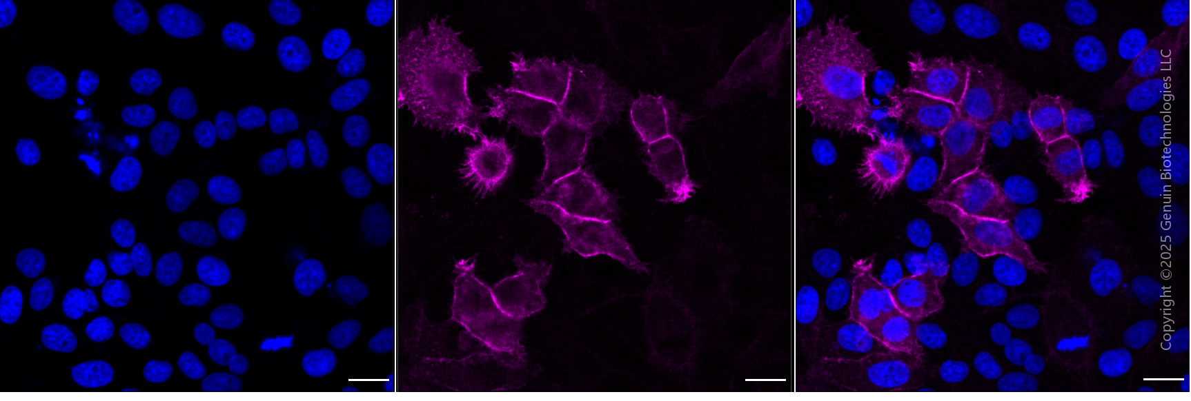
| WB, FC, ICC |
|---|---|
| Primary Accession | P00533 |
| Reactivity | Human |
| Clonality | Monoclonal |
| Isotype | Mouse IgG2b |
| Clone Names | 24GB5795 |
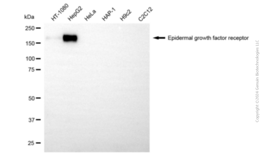
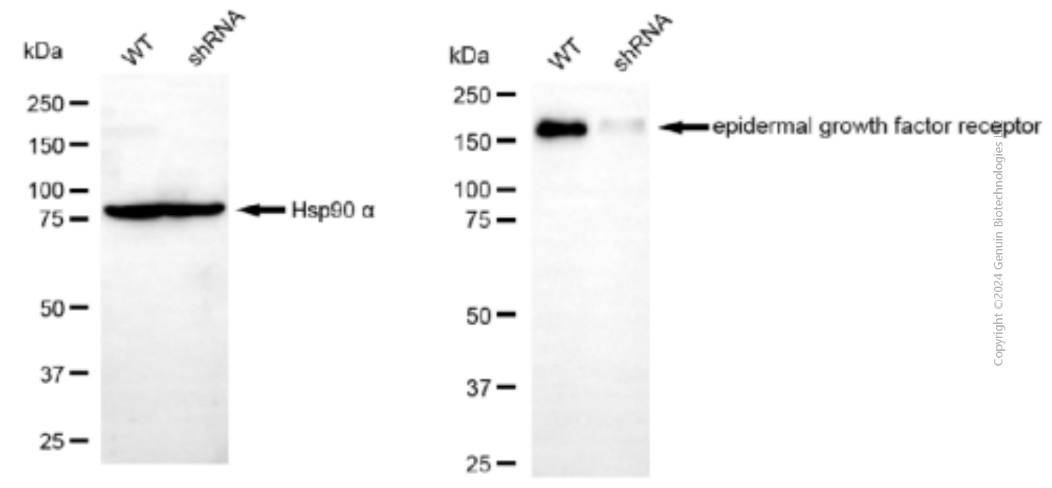
| Calculated MW | Predicted, 134 kDa, Observed, 170 kDa |
| Gene Name | EGFR |
| Aliases | EGFR; Epidermal Growth Factor Receptor; ERBB1; ERRP; ERBB; Receptor Tyrosine-Protein Kinase ErbB-1; Erb-B2 Receptor Tyrosine Kinase 1; Proto-Oncogene C-ErbB-1; EC 2.7.10.1; HER1; Epidermal Growth Factor Receptor (Avian Erythroblastic Leukemia Viral (V-Erb-B) Oncogene Homolog); Erythroblastic Leukemia Viral (V-Erb-B) Oncogene Homolog (Avian); Avian Erythroblastic Leukemia Viral (V-Erb-B) Oncogene Homolog; Epidermal Growth Factor Receptor Tyrosine Kinase Domain; Cell Proliferation-Inducing Protein 61; Cell Growth Inhibiting Protein 40; EGFR VIII; EC 2.7.10; NISBD2; PIG61; MENA |
| Immunogen | Recombinant protein of human EGFR |
| Gene ID | 1956 |
|---|---|
| Other Names | Epidermal growth factor receptor, 2.7.10.1, Proto-oncogene c-ErbB-1, Receptor tyrosine-protein kinase erbB-1, EGFR (HGNC:3236), ERBB, ERBB1, HER1 |
| Name | EGFR (HGNC:3236) |
|---|---|
| Synonyms | ERBB, ERBB1, HER1 |
| Function | Receptor tyrosine kinase binding ligands of the EGF family and activating several signaling cascades to convert extracellular cues into appropriate cellular responses (PubMed:10805725, PubMed:27153536, PubMed:2790960, PubMed:35538033). Known ligands include EGF, TGFA/TGF- alpha, AREG, epigen/EPGN, BTC/betacellulin, epiregulin/EREG and HBEGF/heparin-binding EGF (PubMed:12297049, PubMed:15611079, PubMed:17909029, PubMed:20837704, PubMed:27153536, PubMed:2790960, PubMed:7679104, PubMed:8144591, PubMed:9419975). Ligand binding triggers receptor homo- and/or heterodimerization and autophosphorylation on key cytoplasmic residues. The phosphorylated receptor recruits adapter proteins like GRB2 which in turn activates complex downstream signaling cascades. Activates at least 4 major downstream signaling cascades including the RAS-RAF-MEK-ERK, PI3 kinase-AKT, PLCgamma-PKC and STATs modules (PubMed:27153536). May also activate the NF-kappa-B signaling cascade (PubMed:11116146). Also directly phosphorylates other proteins like RGS16, activating its GTPase activity and probably coupling the EGF receptor signaling to the G protein-coupled receptor signaling (PubMed:11602604). Also phosphorylates MUC1 and increases its interaction with SRC and CTNNB1/beta-catenin (PubMed:11483589). Positively regulates cell migration via interaction with CCDC88A/GIV which retains EGFR at the cell membrane following ligand stimulation, promoting EGFR signaling which triggers cell migration (PubMed:20462955). Plays a role in enhancing learning and memory performance (By similarity). Plays a role in mammalian pain signaling (long-lasting hypersensitivity) (By similarity). |
| Cellular Location | Cell membrane; Single-pass type I membrane protein. Endoplasmic reticulum membrane; Single-pass type I membrane protein Golgi apparatus membrane; Single-pass type I membrane protein. Nucleus membrane; Single-pass type I membrane protein. Endosome. Endosome membrane. Nucleus. Note=In response to EGF, translocated from the cell membrane to the nucleus via Golgi and ER (PubMed:17909029, PubMed:20674546). Endocytosed upon activation by ligand (PubMed:17182860, PubMed:17909029, PubMed:27153536, PubMed:2790960). Colocalized with GPER1 in the nucleus of estrogen agonist-induced cancer-associated fibroblasts (CAF) (PubMed:20551055) |
| Tissue Location | Ubiquitously expressed. Isoform 2 is also expressed in ovarian cancers. |
Research Areas
Citations (0)

Thousands of laboratories across the world have published research that depended on the performance of antibodies from Abcepta to advance their research. Check out links to articles that cite our products in major peer-reviewed journals, organized by research category.
Submit your citation using an Abcepta antibody to
info@abcepta.com, and receive a free "I Love Antibodies" mug.
info@abcepta.com, and receive a free "I Love Antibodies" mug.

Application Protocols
Provided below are standard protocols that you may find useful for product applications.
Abcepta welcomes feedback from its customers.
If you have used an Abcepta product and would like to share how it has performed, please click on the "Submit Review" button and provide the requested information. Our staff will examine and post your review and contact you if needed.
If you have any additional inquiries please email technical services at tech@abcepta.com.
$ 399.20

$ 149.00
Cat# AGI1920
Ordering Information
United States
AlbaniaAustraliaAustriaBelgiumBosnia & HerzegovinaBrazilBulgariaCanadaCentral AmericaChinaCroatiaCyprusCzech RepublicDenmarkEstoniaFinlandFranceGermanyGreeceHong KongHungaryIcelandIndiaIndonesiaIrelandIsraelItalyJapanLatviaLithuaniaLuxembourgMacedoniaMalaysiaMaltaMexicoNetherlandsNew ZealandNorwayPakistanPolandPortugalRomaniaSerbiaSingaporeSlovakiaSloveniaSouth AfricaSouth KoreaSpainSwedenSwitzerlandTaiwanTurkeyUnited KingdomUnited StatesVietnamWorldwideOthers
USA Headquarters
(888) 735-7227 / (858) 622-0099 or (858) 875-1900
Other Products
Shipping Information
Domestic orders (in stock items)
Shipped out the same day. Orders placed after 1 PM (PST) will ship out the next business day.
International orders
Contact your local distributors
