


 Foundational characteristics of cancer include proliferation, angiogenesis, migration, evasion of apoptosis, and cellular immortality. Find key markers for these cellular processes and antibodies to detect them.
 The SUMOplot™ Analysis Program predicts and scores sumoylation sites in your protein. SUMOylation is a post-translational modification involved in various cellular processes, such as nuclear-cytosolic transport, transcriptional regulation, apoptosis, protein stability, response to stress, and progression through the cell cycle.
 The Autophagy Receptor Motif Plotter predicts and scores autophagy receptor binding sites in your protein. Identifying proteins connected to this pathway is critical to understanding the role of autophagy in physiological as well as pathological processes such as development, differentiation, neurodegenerative diseases, stress, infection, and cancer.
> home > Products > Primary Antibodies > Antibody Collections > KD-Validated Antibodies > KD-Validated Anti-Annexin A1 Mouse Monoclonal Antibody
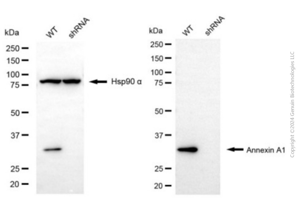
KD-Validated Anti-Annexin A1 Mouse Monoclonal Antibody
Mouse monoclonal antibody
- SPECIFICATION
- CITATIONS
- PROTOCOLS
- BACKGROUND

Application
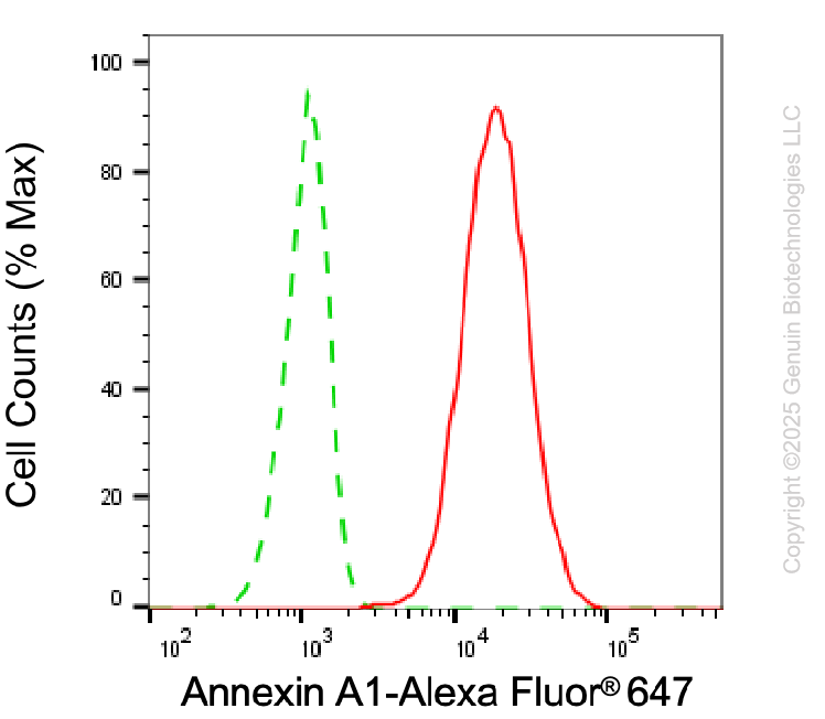
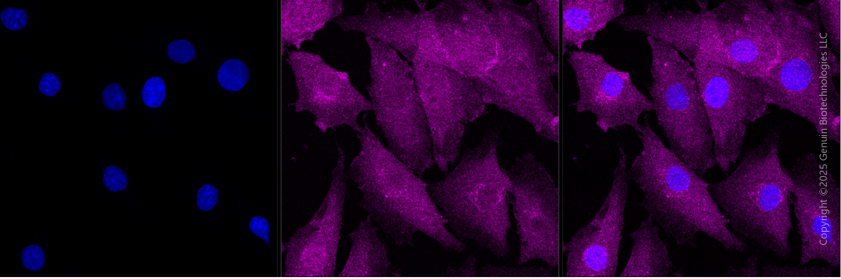
| WB, FC, ICC |
|---|---|
| Primary Accession | P04083 |
| Reactivity | Rat, Human |
| Clonality | Monoclonal |
| Isotype | Mouse IgG1 |
| Clone Names | 24GB6230 |
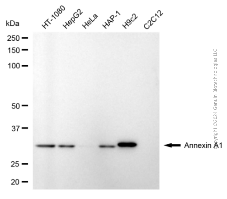
| Calculated MW | Predicted, 39 kDa, observed, 33 kDa |
| Gene Name | ANXA1 |
| Aliases | ANXA1; Annexin A1; ANX1; LPC1; Phospholipase A2 Inhibitory Protein; Chromobindin-9; Calpactin II; Calpactin-2; Annexin-1; Epididymis Secretory Sperm Binding Protein; Annexin I (Lipocortin I); Lipocortin I; Annexin I; P35 |
| Immunogen | Recombinant protein of human ANXA1 |
| Gene ID | 301 |
|---|---|
| Other Names | Annexin A1, Annexin I, Annexin-1, Calpactin II, Calpactin-2, Chromobindin-9, Lipocortin I, Phospholipase A2 inhibitory protein, p35, Annexin Ac2-26, ANXA1, ANX1, LPC1 |
| Name | ANXA1 |
|---|---|
| Synonyms | ANX1, LPC1 |
| Function | Plays important roles in the innate immune response as effector of glucocorticoid-mediated responses and regulator of the inflammatory process. Has anti-inflammatory activity (PubMed:8425544). Plays a role in glucocorticoid-mediated down-regulation of the early phase of the inflammatory response (By similarity). Contributes to the adaptive immune response by enhancing signaling cascades that are triggered by T-cell activation, regulates differentiation and proliferation of activated T-cells (PubMed:17008549). Promotes the differentiation of T-cells into Th1 cells and negatively regulates differentiation into Th2 cells (PubMed:17008549). Has no effect on unstimulated T cells (PubMed:17008549). Negatively regulates hormone exocytosis via activation of the formyl peptide receptors and reorganization of the actin cytoskeleton (PubMed:19625660). Has high affinity for Ca(2+) and can bind up to eight Ca(2+) ions (By similarity). Displays Ca(2+)-dependent binding to phospholipid membranes (PubMed:2532504, PubMed:8557678). Plays a role in the formation of phagocytic cups and phagosomes. Plays a role in phagocytosis by mediating the Ca(2+)-dependent interaction between phagosomes and the actin cytoskeleton (By similarity). |
| Cellular Location | Nucleus. Cytoplasm. Cell projection, cilium {ECO:0000250|UniProtKB:P46193}. Cell membrane. Membrane; Peripheral membrane protein. Endosome membrane {ECO:0000250|UniProtKB:P07150}; Peripheral membrane protein {ECO:0000250|UniProtKB:P07150}. Basolateral cell membrane {ECO:0000250|UniProtKB:P51662}. Apical cell membrane {ECO:0000250|UniProtKB:P10107}. Lateral cell membrane {ECO:0000250|UniProtKB:P10107}. Secreted. Secreted, extracellular space. Cell membrane; Peripheral membrane protein; Extracellular side. Secreted, extracellular exosome. Cytoplasmic vesicle, secretory vesicle lumen. Cell projection, phagocytic cup {ECO:0000250|UniProtKB:P10107}. Early endosome {ECO:0000250|UniProtKB:P19619}. Cytoplasmic vesicle membrane {ECO:0000250|UniProtKB:P19619}; Peripheral membrane protein {ECO:0000250|UniProtKB:P19619}. Note=Secreted, at least in part via exosomes and other secretory vesicles. Detected in exosomes and other extracellular vesicles (PubMed:25664854). Alternatively, the secretion is dependent on protein unfolding and facilitated by the cargo receptor TMED10; it results in the protein translocation from the cytoplasm into ERGIC (endoplasmic reticulum-Golgi intermediate compartment) followed by vesicle entry and secretion (PubMed:32272059). Detected in gelatinase granules in resting neutrophils (PubMed:10772777). Secretion is increased in response to wounding and inflammation (PubMed:25664854). Secretion is increased upon T-cell activation (PubMed:17008549). Neutrophil adhesion to endothelial cells stimulates secretion via gelatinase granules, but foreign particle phagocytosis has no effect (PubMed:10772777). Colocalizes with actin fibers at phagocytic cups (By similarity). Displays calcium-dependent binding to phospholipid membranes (PubMed:2532504, PubMed:8557678) {ECO:0000250|UniProtKB:P10107, ECO:0000269|PubMed:10772777, ECO:0000269|PubMed:17008549, ECO:0000269|PubMed:2532504, ECO:0000269|PubMed:25664854, ECO:0000269|PubMed:32272059, ECO:0000269|PubMed:8557678} |
| Tissue Location | Detected in resting neutrophils (PubMed:10772777). Detected in peripheral blood T-cells (PubMed:17008549). Detected in extracellular vesicles in blood serum from patients with inflammatory bowel disease, but not in serum from healthy donors (PubMed:25664854) Detected in placenta (at protein level) (PubMed:2532504). Detected in liver. |
Research Areas
Citations (0)

Thousands of laboratories across the world have published research that depended on the performance of antibodies from Abcepta to advance their research. Check out links to articles that cite our products in major peer-reviewed journals, organized by research category.
Submit your citation using an Abcepta antibody to
info@abcepta.com, and receive a free "I Love Antibodies" mug.
info@abcepta.com, and receive a free "I Love Antibodies" mug.

Application Protocols
Provided below are standard protocols that you may find useful for product applications.
Abcepta welcomes feedback from its customers.
If you have used an Abcepta product and would like to share how it has performed, please click on the "Submit Review" button and provide the requested information. Our staff will examine and post your review and contact you if needed.
If you have any additional inquiries please email technical services at tech@abcepta.com.
$ 399.20

$ 149.00
Cat# AGI1929
Ordering Information
United States
AlbaniaAustraliaAustriaBelgiumBosnia & HerzegovinaBrazilBulgariaCanadaCentral AmericaChinaCroatiaCyprusCzech RepublicDenmarkEstoniaFinlandFranceGermanyGreeceHong KongHungaryIcelandIndiaIndonesiaIrelandIsraelItalyJapanLatviaLithuaniaLuxembourgMacedoniaMalaysiaMaltaMexicoNetherlandsNew ZealandNorwayPakistanPolandPortugalRomaniaSerbiaSingaporeSlovakiaSloveniaSouth AfricaSouth KoreaSpainSwedenSwitzerlandTaiwanTurkeyUnited KingdomUnited StatesVietnamWorldwideOthers
USA Headquarters
(888) 735-7227 / (858) 622-0099 or (858) 875-1900
Other Products
Shipping Information
Domestic orders (in stock items)
Shipped out the same day. Orders placed after 1 PM (PST) will ship out the next business day.
International orders
Contact your local distributors
