


 Foundational characteristics of cancer include proliferation, angiogenesis, migration, evasion of apoptosis, and cellular immortality. Find key markers for these cellular processes and antibodies to detect them.
 The SUMOplot™ Analysis Program predicts and scores sumoylation sites in your protein. SUMOylation is a post-translational modification involved in various cellular processes, such as nuclear-cytosolic transport, transcriptional regulation, apoptosis, protein stability, response to stress, and progression through the cell cycle.
 The Autophagy Receptor Motif Plotter predicts and scores autophagy receptor binding sites in your protein. Identifying proteins connected to this pathway is critical to understanding the role of autophagy in physiological as well as pathological processes such as development, differentiation, neurodegenerative diseases, stress, infection, and cancer.
> home > Products > Primary Antibodies > Antibody Collections > KD-Validated Antibodies > KD-Validated Anti-RAB27 Mouse Monoclonal Antibody
KD-Validated Anti-RAB27 Mouse Monoclonal Antibody
Mouse monoclonal antibody
- SPECIFICATION
- CITATIONS
- PROTOCOLS
- BACKGROUND

Application
| WB |
|---|---|
| Primary Accession | P51159 |
| Reactivity | Human |
| Clonality | Monoclonal |
| Isotype | Mouse IgG1 kappa |
| Clone Names | 24GB7165 |
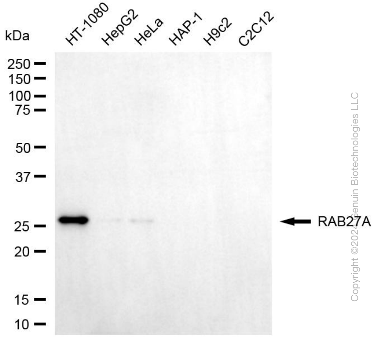
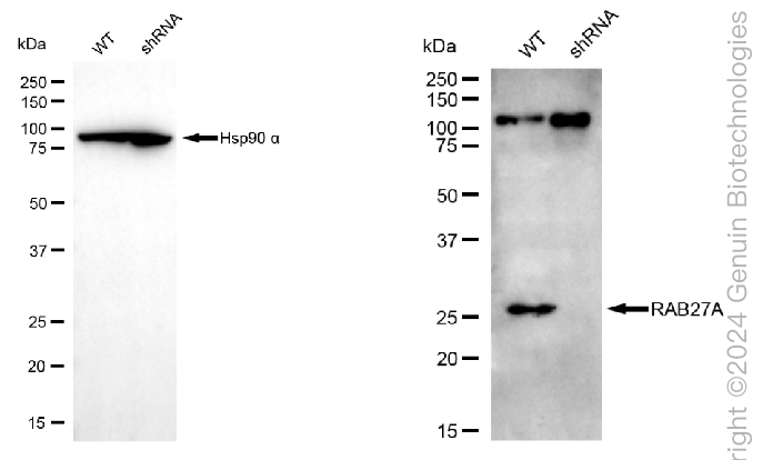
| Calculated MW | Predicted, 25 kDa, observed, 27 kDa |
| Gene Name | RAB27A |
| Aliases | RAB27A; RAB27A, Member RAS Oncogene Family; RAB27; HsT18676; RAM; GS2; Ras-Related Protein Rab-27A; GTP-Binding Protein Ram; Rab-27; Mutant Ras-Related Protein Rab-27A; EC 3.6.5.2 |
| Immunogen | Recombinant protein of human RAB27 |
| Gene ID | 5873 |
|---|---|
| Other Names | Ras-related protein Rab-27A, Rab-27, 3.6.5.2, GTP-binding protein Ram, RAB27A, RAB27 |
| Name | RAB27A |
|---|---|
| Synonyms | RAB27 |
| Function | Small GTPase which cycles between active GTP-bound and inactive GDP-bound states. In its active state, binds to a variety of effector proteins to regulate homeostasis of late endocytic pathway, including endosomal positioning, maturation and secretion (PubMed:30771381). Plays a role in cytotoxic granule exocytosis in lymphocytes. Required for both granule maturation and granule docking and priming at the immunologic synapse. |
| Cellular Location | Membrane; Lipid-anchor. Melanosome. Late endosome. Lysosome. Note=Identified by mass spectrometry in melanosome fractions from stage I to stage IV (PubMed:12643545, PubMed:17081065). Localizes to endosomal exocytic vesicles (PubMed:17237785). |
| Tissue Location | Found in all the examined tissues except in brain. Low expression was found in thymus, kidney, muscle and placenta Detected in melanocytes, and in most tumor cell lines examined Expressed in cytotoxic T-lymphocytes (CTL) and mast cells |
Research Areas
Citations (0)

Thousands of laboratories across the world have published research that depended on the performance of antibodies from Abcepta to advance their research. Check out links to articles that cite our products in major peer-reviewed journals, organized by research category.
Submit your citation using an Abcepta antibody to
info@abcepta.com, and receive a free "I Love Antibodies" mug.
info@abcepta.com, and receive a free "I Love Antibodies" mug.

Application Protocols
Provided below are standard protocols that you may find useful for product applications.
Abcepta welcomes feedback from its customers.
If you have used an Abcepta product and would like to share how it has performed, please click on the "Submit Review" button and provide the requested information. Our staff will examine and post your review and contact you if needed.
If you have any additional inquiries please email technical services at tech@abcepta.com.
$ 399.20

$ 149.00
Cat# AGI1995
Ordering Information
United States
AlbaniaAustraliaAustriaBelgiumBosnia & HerzegovinaBrazilBulgariaCanadaCentral AmericaChinaCroatiaCyprusCzech RepublicDenmarkEstoniaFinlandFranceGermanyGreeceHong KongHungaryIcelandIndiaIndonesiaIrelandIsraelItalyJapanLatviaLithuaniaLuxembourgMacedoniaMalaysiaMaltaMexicoNetherlandsNew ZealandNorwayPakistanPolandPortugalRomaniaSerbiaSingaporeSlovakiaSloveniaSouth AfricaSouth KoreaSpainSwedenSwitzerlandTaiwanTurkeyUnited KingdomUnited StatesVietnamWorldwideOthers
USA Headquarters
(888) 735-7227 / (858) 622-0099 or (858) 875-1900
Other Products
Shipping Information
Domestic orders (in stock items)
Shipped out the same day. Orders placed after 1 PM (PST) will ship out the next business day.
International orders
Contact your local distributors
