


 Foundational characteristics of cancer include proliferation, angiogenesis, migration, evasion of apoptosis, and cellular immortality. Find key markers for these cellular processes and antibodies to detect them.
 The SUMOplot™ Analysis Program predicts and scores sumoylation sites in your protein. SUMOylation is a post-translational modification involved in various cellular processes, such as nuclear-cytosolic transport, transcriptional regulation, apoptosis, protein stability, response to stress, and progression through the cell cycle.
 The Autophagy Receptor Motif Plotter predicts and scores autophagy receptor binding sites in your protein. Identifying proteins connected to this pathway is critical to understanding the role of autophagy in physiological as well as pathological processes such as development, differentiation, neurodegenerative diseases, stress, infection, and cancer.
> home > Products > Primary Antibodies > Antibody Collections > KD-Validated Antibodies > KD-Validated Anti-Optineurin Rabbit Monoclonal Antibody
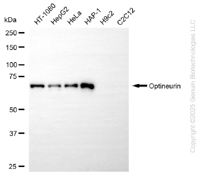
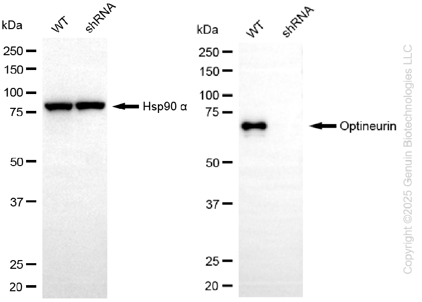
KD-Validated Anti-Optineurin Rabbit Monoclonal Antibody
Rabbit monoclonal antibody
- SPECIFICATION
- CITATIONS
- PROTOCOLS
- BACKGROUND

Application
| WB |
|---|---|
| Primary Accession | Q96CV9 |
| Reactivity | Human |
| Clonality | Monoclonal |
| Isotype | Rabbit IgG |
| Clone Names | 25GB4715 |
| Calculated MW | Predicted, 66 kDa; observed, 70 kDa |
| Gene Name | OPTN |
| Aliases | OPTN; Optineurin; FIP-2; FIP2; HYPL; HIP7; NRP; TFIIIA-INTP; GLC1E; Transcription Factor IIIA-Interacting Protein; Optic Neuropathy-Inducing Protein; Huntingtin-Interacting Protein 7; Huntingtin-Interacting Protein L; E3-14.7K-Interacting Protein; Huntingtin Yeast Partner L; HIP-7; Tumor Necrosis Factor Alpha-Inducible Cellular Protein Containing Leucine Zipper Domains; Transcrption Factor IIIA-Interacting Protein; Glaucoma 1, Open Angle, E (Adult-Onset); Huntingtin Interacting Protein L; Nemo-Related Protein; NEMO-Related Protein; TFIIIA-IntP; ALS12 |
| Immunogen | Recombinant protein of human Optineurin |
| Gene ID | 10133 |
|---|---|
| Other Names | Optineurin, E3-14.7K-interacting protein, FIP-2, Huntingtin yeast partner L, Huntingtin-interacting protein 7, HIP-7, Huntingtin-interacting protein L, NEMO-related protein, Optic neuropathy-inducing protein, Transcription factor IIIA-interacting protein, TFIIIA-IntP, OPTN |
| Name | OPTN |
|---|---|
| Function | Plays an important role in the maintenance of the Golgi complex, in membrane trafficking, in exocytosis, through its interaction with myosin VI and Rab8 (PubMed:27534431). Links myosin VI to the Golgi complex and plays an important role in Golgi ribbon formation (PubMed:27534431). Plays a role in the activation of innate immune response during viral infection. Mechanistically, recruits TBK1 at the Golgi apparatus, promoting its trans-phosphorylation after RLR or TLR3 stimulation (PubMed:27538435). In turn, activated TBK1 phosphorylates its downstream partner IRF3 to produce IFN-beta/IFNB1. Plays a neuroprotective role in the eye and optic nerve. May act by regulating membrane trafficking and cellular morphogenesis via a complex that contains Rab8 and huntingtin (HD). Mediates the interaction of Rab8 with the probable GTPase-activating protein TBC1D17 during Rab8-mediated endocytic trafficking, such as that of transferrin receptor (TFRC/TfR); regulates Rab8 recruitment to tubules emanating from the endocytic recycling compartment (PubMed:22854040). Autophagy receptor that interacts directly with both the cargo to become degraded and an autophagy modifier of the MAP1 LC3 family; targets ubiquitin- coated bacteria (xenophagy), such as cytoplasmic Salmonella enterica, and appears to function in the same pathway as SQSTM1 and CALCOCO2/NDP52. |
| Cellular Location | Cytoplasm, perinuclear region. Golgi apparatus. Golgi apparatus, trans-Golgi network Cytoplasmic vesicle, autophagosome. Cytoplasmic vesicle. Recycling endosome. Note=Found in the perinuclear region and associates with the Golgi apparatus (PubMed:27534431) Colocalizes with MYO6 and RAB8 at the Golgi complex and in vesicular structures close to the plasma membrane. Localizes to LC3-positive cytoplasmic vesicles upon induction of autophagy |
| Tissue Location | Present in aqueous humor of the eye (at protein level). Expressed in the trabecular meshwork (at protein level) (PubMed:11834836, PubMed:12379221, PubMed:12646749). Expressed in nonpigmented ciliary epithelium (at protein level) (PubMed:11834836) Expressed at high levels in skeletal muscle, also detected in heart, brain, pancreas, kidney, placenta and liver (PubMed:9488477). Expressed in dermal fibroblasts (at protein level) (PubMed:11834836) |
Research Areas
Citations (0)

Thousands of laboratories across the world have published research that depended on the performance of antibodies from Abcepta to advance their research. Check out links to articles that cite our products in major peer-reviewed journals, organized by research category.
Submit your citation using an Abcepta antibody to
info@abcepta.com, and receive a free "I Love Antibodies" mug.
info@abcepta.com, and receive a free "I Love Antibodies" mug.

Application Protocols
Provided below are standard protocols that you may find useful for product applications.
Abcepta welcomes feedback from its customers.
If you have used an Abcepta product and would like to share how it has performed, please click on the "Submit Review" button and provide the requested information. Our staff will examine and post your review and contact you if needed.
If you have any additional inquiries please email technical services at tech@abcepta.com.
$ 399.20

$ 149.00
Cat# AGI2284
Ordering Information
United States
AlbaniaAustraliaAustriaBelgiumBosnia & HerzegovinaBrazilBulgariaCanadaCentral AmericaChinaCroatiaCyprusCzech RepublicDenmarkEstoniaFinlandFranceGermanyGreeceHong KongHungaryIcelandIndiaIndonesiaIrelandIsraelItalyJapanLatviaLithuaniaLuxembourgMacedoniaMalaysiaMaltaMexicoNetherlandsNew ZealandNorwayPakistanPolandPortugalRomaniaSerbiaSingaporeSlovakiaSloveniaSouth AfricaSouth KoreaSpainSwedenSwitzerlandTaiwanTurkeyUnited KingdomUnited StatesVietnamWorldwideOthers
USA Headquarters
(888) 735-7227 / (858) 622-0099 or (858) 875-1900
Other Products
Shipping Information
Domestic orders (in stock items)
Shipped out the same day. Orders placed after 1 PM (PST) will ship out the next business day.
International orders
Contact your local distributors
