


 Foundational characteristics of cancer include proliferation, angiogenesis, migration, evasion of apoptosis, and cellular immortality. Find key markers for these cellular processes and antibodies to detect them.
 The SUMOplot™ Analysis Program predicts and scores sumoylation sites in your protein. SUMOylation is a post-translational modification involved in various cellular processes, such as nuclear-cytosolic transport, transcriptional regulation, apoptosis, protein stability, response to stress, and progression through the cell cycle.
 The Autophagy Receptor Motif Plotter predicts and scores autophagy receptor binding sites in your protein. Identifying proteins connected to this pathway is critical to understanding the role of autophagy in physiological as well as pathological processes such as development, differentiation, neurodegenerative diseases, stress, infection, and cancer.
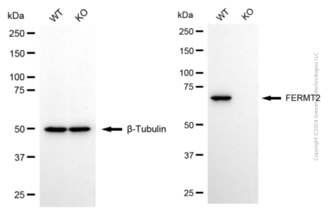
> home > Products > Primary Antibodies > Antibody Collections > KD-Validated Antibodies > KO-Validated Anti-FERMT2 Mouse Monoclonal Antibody
KO-Validated Anti-FERMT2 Mouse Monoclonal Antibody
Mouse monoclonal antibody
- SPECIFICATION
- CITATIONS
- PROTOCOLS
- BACKGROUND

Application
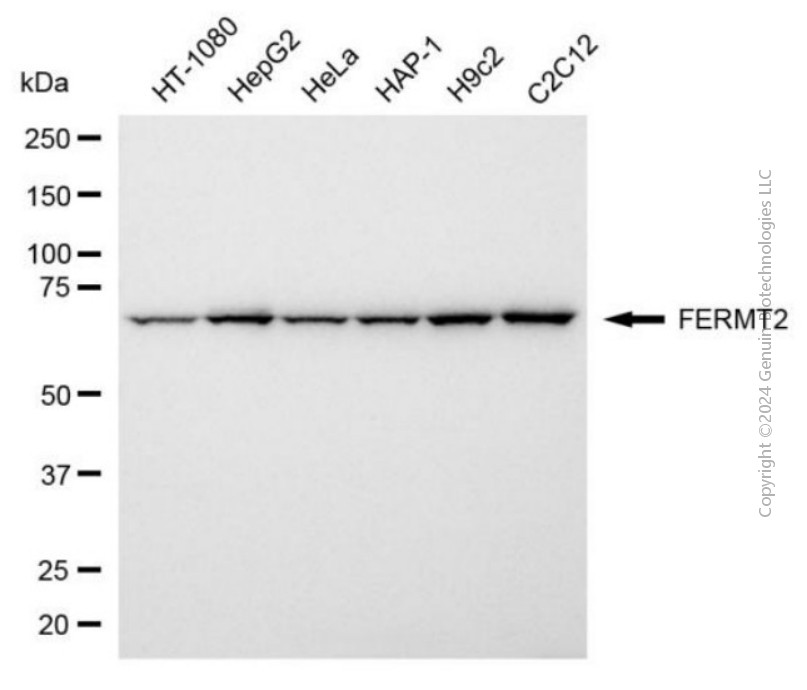
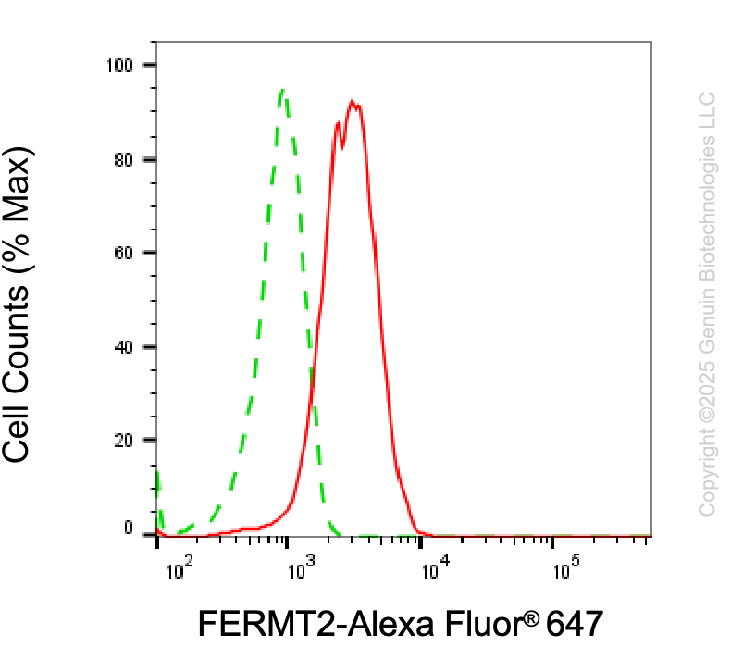
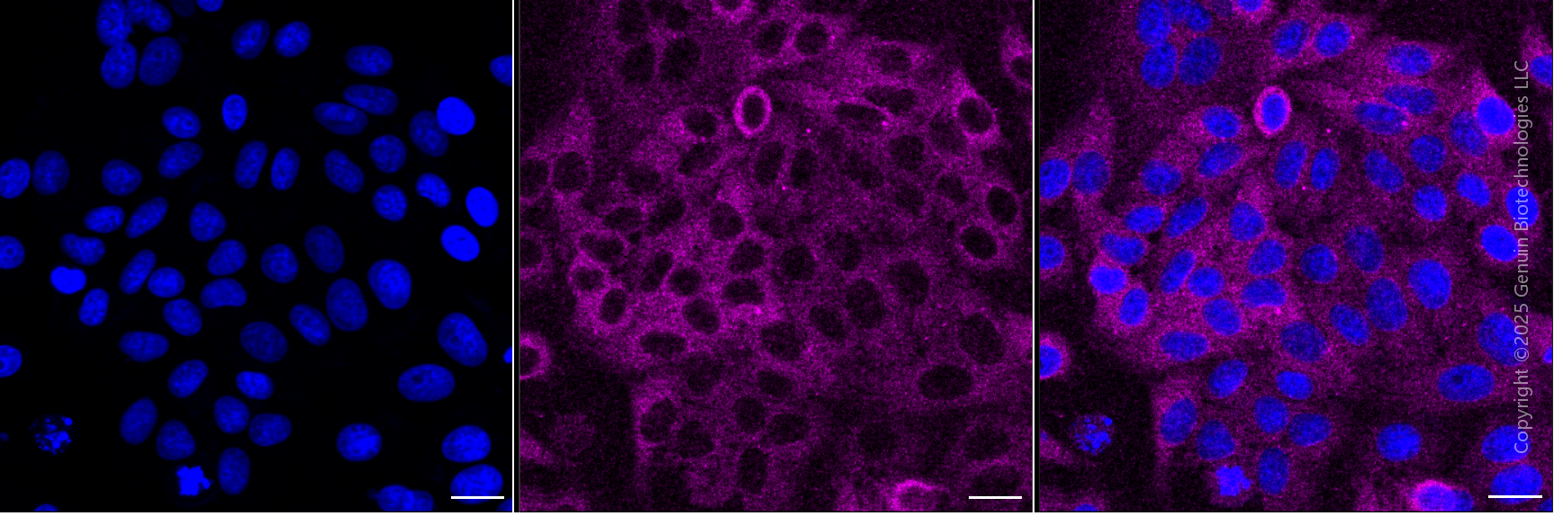
| WB, FC, ICC |
|---|---|
| Primary Accession | Q96AC1 |
| Reactivity | Rat, Human, Mouse |
| Clonality | Monoclonal |
| Isotype | Mouse IgG2a |
| Clone Names | 24GB6195 |
| Calculated MW | Predicted, 78 kDa, observed, 73 kDa |
| Gene Name | FERMT2 |
| Aliases | FERMT2; FERM Domain Containing Kindlin 2; KIND2; UNC112B; PLEKHC1; Mig-2; Pleckstrin Homology Domain Containing, Family C (With FERM Domain) Member 1; PH Domain-Containing Family C Member 1; Fermitin Family Homolog 2; Mitogen Inducible Gene-2; Fermitin Family Member 2; Kindlin-2; MIG2; Pleckstrin Homology Domain Containing, Family C Member 1; Pleckstrin Homology Domain-Containing Family C Member 1; Fermitin Family Homolog 2 (Drosophila); Mitogen Inducible Gene 2 Protein; Mitogen-Inducible Gene 2 Protein; Kindlin 2; UNC112; MIG-2 |
| Immunogen | Recombinant protein of human FERMT2 |
| Gene ID | 10979 |
|---|---|
| Other Names | Fermitin family homolog 2, Kindlin-2, Mitogen-inducible gene 2 protein, MIG-2, Pleckstrin homology domain-containing family C member 1, PH domain-containing family C member 1, FERMT2, KIND2, MIG2, PLEKHC1 |
| Name | FERMT2 |
|---|---|
| Synonyms | KIND2, MIG2, PLEKHC1 |
| Function | Scaffolding protein that enhances integrin activation mediated by TLN1 and/or TLN2, but activates integrins only weakly by itself. Binds to membranes enriched in phosphoinositides. Enhances integrin-mediated cell adhesion onto the extracellular matrix and cell spreading; this requires both its ability to interact with integrins and with phospholipid membranes. Required for the assembly of focal adhesions. Participates in the connection between extracellular matrix adhesion sites and the actin cytoskeleton and also in the orchestration of actin assembly and cell shape modulation. Recruits FBLIM1 to focal adhesions. Plays a role in the TGFB1 and integrin signaling pathways. Stabilizes active CTNNB1 and plays a role in the regulation of transcription mediated by CTNNB1 and TCF7L2/TCF4 and in Wnt signaling. |
| Cellular Location | Cytoplasm. Cytoplasm, cell cortex. Cytoplasm, cytoskeleton. Cytoplasm, cytoskeleton, stress fiber. Cell junction, focal adhesion. Membrane; Peripheral membrane protein; Cytoplasmic side. Cell projection, lamellipodium membrane; Peripheral membrane protein; Cytoplasmic side. Nucleus. Cytoplasm, myofibril, sarcomere, I band. Cell surface. Note=Colocalizes with actin stress fibers at cell-ECM focal adhesion sites. Colocalizes with ITGB3 at lamellipodia at the leading edge of spreading cells. Binds to membranes that contain phosphatidylinositides |
| Tissue Location | Ubiquitous. Found in numerous tumor tissues. |
Research Areas
Citations (0)

Thousands of laboratories across the world have published research that depended on the performance of antibodies from Abcepta to advance their research. Check out links to articles that cite our products in major peer-reviewed journals, organized by research category.
Submit your citation using an Abcepta antibody to
info@abcepta.com, and receive a free "I Love Antibodies" mug.
info@abcepta.com, and receive a free "I Love Antibodies" mug.

Application Protocols
Provided below are standard protocols that you may find useful for product applications.
Abcepta welcomes feedback from its customers.
If you have used an Abcepta product and would like to share how it has performed, please click on the "Submit Review" button and provide the requested information. Our staff will examine and post your review and contact you if needed.
If you have any additional inquiries please email technical services at tech@abcepta.com.
$ 149.00
$ 499.00
Cat# AGI2401
Ordering Information
United States
AlbaniaAustraliaAustriaBelgiumBosnia & HerzegovinaBrazilBulgariaCanadaCentral AmericaChinaCroatiaCyprusCzech RepublicDenmarkEstoniaFinlandFranceGermanyGreeceHong KongHungaryIcelandIndiaIndonesiaIrelandIsraelItalyJapanLatviaLithuaniaLuxembourgMacedoniaMalaysiaMaltaMexicoNetherlandsNew ZealandNorwayPakistanPolandPortugalRomaniaSerbiaSingaporeSlovakiaSloveniaSouth AfricaSouth KoreaSpainSwedenSwitzerlandTaiwanTurkeyUnited KingdomUnited StatesVietnamWorldwideOthers
USA Headquarters
(888) 735-7227 / (858) 622-0099 or (858) 875-1900
Other Products
Shipping Information
Domestic orders (in stock items)
Shipped out the same day. Orders placed after 1 PM (PST) will ship out the next business day.
International orders
Contact your local distributors
