


 Foundational characteristics of cancer include proliferation, angiogenesis, migration, evasion of apoptosis, and cellular immortality. Find key markers for these cellular processes and antibodies to detect them.
 The SUMOplot™ Analysis Program predicts and scores sumoylation sites in your protein. SUMOylation is a post-translational modification involved in various cellular processes, such as nuclear-cytosolic transport, transcriptional regulation, apoptosis, protein stability, response to stress, and progression through the cell cycle.
 The Autophagy Receptor Motif Plotter predicts and scores autophagy receptor binding sites in your protein. Identifying proteins connected to this pathway is critical to understanding the role of autophagy in physiological as well as pathological processes such as development, differentiation, neurodegenerative diseases, stress, infection, and cancer.
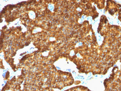
Chromogranin A / CHGA (Neuroendocrine Marker) Antibody - With BSA and Azide
Mouse Monoclonal Antibody [Clone SPM553 ]
- SPECIFICATION
- CITATIONS
- PROTOCOLS
- BACKGROUND

Application
| IHC-P, IF, FC |
|---|---|
| Primary Accession | P10645 |
| Other Accession | 1113, 150793 |
| Reactivity | Human |
| Host | Mouse |
| Clonality | Monoclonal |
| Isotype | Mouse / IgG1, kappa |
| Clone Names | SPM553 |
| Calculated MW | 68-75kDa |
| Gene ID | 1113 |
|---|---|
| Other Names | Chromogranin-A, CgA, Pituitary secretory protein I, SP-I, Vasostatin-1, Vasostatin I, Vasostatin-2, Vasostatin II, EA-92, ES-43, Pancreastatin, SS-18, WA-8, WE-14, LF-19, AL-11, GV-19, GR-44, ER-37, CHGA |
| Application Note | IHC-P~~N/A IF~~1:50~200 FC~~1:10~50 |
| Format | 200ug/ml of Ab purified from Bioreactor Concentrate by Protein A/G. Prepared in 10mM PBS with 0.05% BSA & 0.05% azide. Also available WITHOUT BSA & azide at 1.0mg/ml. |
| Storage | Store at 2 to 8°C.Antibody is stable for 24 months. |
| Precautions | Chromogranin A / CHGA (Neuroendocrine Marker) Antibody - With BSA and Azide is for research use only and not for use in diagnostic or therapeutic procedures. |
| Name | CHGA |
|---|---|
| Function | [Pancreastatin]: Strongly inhibits glucose induced insulin release from the pancreas. [Serpinin]: Regulates granule biogenesis in endocrine cells by up-regulating the transcription of protease nexin 1 (SERPINE2) via a cAMP-PKA-SP1 pathway. This leads to inhibition of granule protein degradation in the Golgi complex which in turn promotes granule formation. |
| Cellular Location | [Serpinin]: Secreted {ECO:0000250|UniProtKB:P26339}. Cytoplasmic vesicle, secretory vesicle {ECO:0000250|UniProtKB:P26339}. Note=Pyroglutaminated serpinin localizes to secretory vesicle. {ECO:0000250|UniProtKB:P26339} |
| Tissue Location | Detected in cerebrospinal fluid (at protein level) (PubMed:25326458). Detected in urine (at protein level) (PubMed:37453717). |

Thousands of laboratories across the world have published research that depended on the performance of antibodies from Abcepta to advance their research. Check out links to articles that cite our products in major peer-reviewed journals, organized by research category.
info@abcepta.com, and receive a free "I Love Antibodies" mug.

Provided below are standard protocols that you may find useful for product applications.
Background
Chromogranin A is present in neuroendocrine cells throughout the body, including the neuroendocrine cells of the large and small intestine, adrenal medulla and pancreatic islets. It is an excellent marker for carcinoid tumors, pheochromocytomas, paragangliomas, and other neuroendocrine tumors. Co-expression of chromogranin A and neuron specific enolase (NSE) is common in neuroendocrine neoplasms. Reportedly, co-expression of certain keratins and chromogranin indicates neuroendocrine lineage. The presence of strong anti-chromogranin staining and absence of anti-keratin staining should raise the possibility of paraganglioma. The co-expression of chromogranin and NSE is typical of neuroendocrine neoplasms. Most pituitary adenomas and prolactinomas readily express chromogranin.
References
Konecki DS et. al. J Biol Chem 1987;262:17026-30. | Lloyd RV et. al. Am J Pathol 1988; 130:296-304
If you have used an Abcepta product and would like to share how it has performed, please click on the "Submit Review" button and provide the requested information. Our staff will examine and post your review and contact you if needed.
If you have any additional inquiries please email technical services at tech@abcepta.com.
Ordering Information
Other Products
Shipping Information
