


 Foundational characteristics of cancer include proliferation, angiogenesis, migration, evasion of apoptosis, and cellular immortality. Find key markers for these cellular processes and antibodies to detect them.
 The SUMOplot™ Analysis Program predicts and scores sumoylation sites in your protein. SUMOylation is a post-translational modification involved in various cellular processes, such as nuclear-cytosolic transport, transcriptional regulation, apoptosis, protein stability, response to stress, and progression through the cell cycle.
 The Autophagy Receptor Motif Plotter predicts and scores autophagy receptor binding sites in your protein. Identifying proteins connected to this pathway is critical to understanding the role of autophagy in physiological as well as pathological processes such as development, differentiation, neurodegenerative diseases, stress, infection, and cancer.
Anti-GP2 (Glycoprotein 2) / ZAP75 Antibody
Mouse Monoclonal Antibody
- SPECIFICATION
- CITATIONS
- PROTOCOLS
- BACKGROUND

Application
| IHC-P, IF, FC |
|---|---|
| Primary Accession | P55259 |
| Other Accession | 53985 |
| Reactivity | Human |
| Host | Mouse |
| Clonality | Monoclonal |
| Isotype | Mouse / IgG |
| Clone Names | GP2/1712 |
| Calculated MW | 59480 Da |
| Gene ID | 2813 |
|---|---|
| Other Names | Glycoprotein 2 (zymogen granule membrane); GP2; Pancreatic zymogen granule membrane associated protein GP2; Pancreatic zymogen granule membrane protein GP-2; ZAP75 |
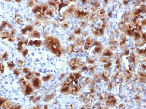
| Application Note | Flow Cytometry (0.5-1ug/million cells); Immunofluorescence (0.5-1ug/ml); ,Immunohistology (Formalin-fixed) (1-2ug/ml for 30 minutes at RT),(Staining of formalin-fixed tissues is enhanced by boiling tissue sections in 10mM Tris with 1mM EDTA, pH 9.0 for 10-20 min followed by cooling at RT for 20 minutes),Optimal dilution for a specific application should be determined. |
| Format | 200ug/ml of Ab purified from Bioreactor Concentrate by Protein A/G. Prepared in 10mM PBS with 0.05% BSA & 0.05% azide. Also available WITHOUT BSA & azide at 1.0mg/ml. |
| Storage | Store at 2 to 8°C.Antibody is stable for 24 months. |
| Precautions | Anti-GP2 (Glycoprotein 2) / ZAP75 Antibody is for research use only and not for use in diagnostic or therapeutic procedures. |
| Name | GP2 (HGNC:4441) |
|---|---|
| Function | Functions as an intestinal M-cell transcytotic receptor specific for type-I-piliated bacteria that participates in the mucosal immune response toward these bacteria. At the apical membrane of M- cells it binds fimH, a protein of the bacteria type I pilus tip. Internalizes bound bacteria, like E.coli and S.typhimurium, from the lumen of the intestine and delivers them, through M-cells, to the underlying organized lymphoid follicles where they are captured by antigen-presenting dendritic cells to elicit a mucosal immune response. |
| Cellular Location | Zymogen granule membrane {ECO:0000250|UniProtKB:P19218}; Lipid-anchor, GPI-anchor {ECO:0000250|UniProtKB:P19218}. Secreted Cell membrane {ECO:0000250|UniProtKB:P19218}; Lipid-anchor, GPI-anchor {ECO:0000250|UniProtKB:P19218}. Apical cell membrane {ECO:0000250|UniProtKB:Q9D733}; Lipid-anchor, GPI-anchor {ECO:0000250|UniProtKB:P19218}. Membrane raft {ECO:0000250|UniProtKB:P19218}; Lipid-anchor, GPI-anchor {ECO:0000250|UniProtKB:P19218}. Endosome {ECO:0000250|UniProtKB:Q9D733}. Note=Secreted, after cleavage, in the pancreatic juice. |
| Tissue Location | Expressed in pancreas (at protein level) (PubMed:10760606, PubMed:8666297). Specifically expressed by M (microfold) cells which are atypical epithelial cells of the intestine (at protein level) (PubMed:19907495). |

Thousands of laboratories across the world have published research that depended on the performance of antibodies from Abcepta to advance their research. Check out links to articles that cite our products in major peer-reviewed journals, organized by research category.
info@abcepta.com, and receive a free "I Love Antibodies" mug.

Provided below are standard protocols that you may find useful for product applications.
Background
GP2 (glycoprotein 2), also known as ZAP75, is a 537 amino acid secreted protein. It is an integral membrane protein that is secreted from intracellular zymogen granules and associates with the plasma membrane via glycosylphosphatidylinositol (GPI) linkage. GP2 is cleaved and then released into the pancreatic duct along with exocrine secretions. GP2 binds pathogens such as enterobacteria, thereby playing an important role in the innate immune response. The C-terminus of this protein is related to the C-terminus of the protein encoded by the neighboring gene, uromodulin (UMOD). GP2 is also expressed on the apical plasma membrane of specialized microfold (M) cells among enterocytes and serves as a transcytotic receptor for mucosal antigens. M cells are considered a promising target for oral vaccination against various infectious diseases.
If you have used an Abcepta product and would like to share how it has performed, please click on the "Submit Review" button and provide the requested information. Our staff will examine and post your review and contact you if needed.
If you have any additional inquiries please email technical services at tech@abcepta.com.
Ordering Information
Other Products
Shipping Information
