


 Foundational characteristics of cancer include proliferation, angiogenesis, migration, evasion of apoptosis, and cellular immortality. Find key markers for these cellular processes and antibodies to detect them.
 The SUMOplot™ Analysis Program predicts and scores sumoylation sites in your protein. SUMOylation is a post-translational modification involved in various cellular processes, such as nuclear-cytosolic transport, transcriptional regulation, apoptosis, protein stability, response to stress, and progression through the cell cycle.
 The Autophagy Receptor Motif Plotter predicts and scores autophagy receptor binding sites in your protein. Identifying proteins connected to this pathway is critical to understanding the role of autophagy in physiological as well as pathological processes such as development, differentiation, neurodegenerative diseases, stress, infection, and cancer.
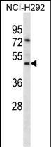
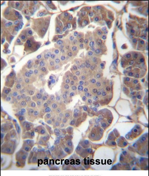
GP2 Antibody (N-term)
Affinity Purified Rabbit Polyclonal Antibody (Pab)
- SPECIFICATION
- CITATIONS
- PROTOCOLS
- BACKGROUND

Application
| IHC-P, WB, E |
|---|---|
| Primary Accession | P55259 |
| Other Accession | NP_001007241.2, NP_001493.2, NP_001007243.2 |
| Reactivity | Human |
| Host | Rabbit |
| Clonality | Polyclonal |
| Isotype | Rabbit IgG |
| Calculated MW | 59480 Da |
| Antigen Region | 55-84 aa |
| Gene ID | 2813 |
|---|---|
| Other Names | Pancreatic secretory granule membrane major glycoprotein GP2, Pancreatic zymogen granule membrane protein GP-2, ZAP75, GP2 |
| Target/Specificity | This GP2 antibody is generated from rabbits immunized with a KLH conjugated synthetic peptide between 55-84 amino acids from the N-terminal region of human GP2. |
| Dilution | IHC-P~~1:10~50 WB~~1:1000 E~~Use at an assay dependent concentration. |
| Format | Purified polyclonal antibody supplied in PBS with 0.09% (W/V) sodium azide. This antibody is purified through a protein A column, followed by peptide affinity purification. |
| Storage | Maintain refrigerated at 2-8°C for up to 2 weeks. For long term storage store at -20°C in small aliquots to prevent freeze-thaw cycles. |
| Precautions | GP2 Antibody (N-term) is for research use only and not for use in diagnostic or therapeutic procedures. |
| Name | GP2 (HGNC:4441) |
|---|---|
| Function | Functions as an intestinal M-cell transcytotic receptor specific for type-I-piliated bacteria that participates in the mucosal immune response toward these bacteria. At the apical membrane of M- cells it binds fimH, a protein of the bacteria type I pilus tip. Internalizes bound bacteria, like E.coli and S.typhimurium, from the lumen of the intestine and delivers them, through M-cells, to the underlying organized lymphoid follicles where they are captured by antigen-presenting dendritic cells to elicit a mucosal immune response. |
| Cellular Location | Zymogen granule membrane {ECO:0000250|UniProtKB:P19218}; Lipid-anchor, GPI-anchor {ECO:0000250|UniProtKB:P19218}. Secreted Cell membrane {ECO:0000250|UniProtKB:P19218}; Lipid-anchor, GPI-anchor {ECO:0000250|UniProtKB:P19218}. Apical cell membrane {ECO:0000250|UniProtKB:Q9D733}; Lipid-anchor, GPI-anchor {ECO:0000250|UniProtKB:P19218}. Membrane raft {ECO:0000250|UniProtKB:P19218}; Lipid-anchor, GPI-anchor {ECO:0000250|UniProtKB:P19218}. Endosome {ECO:0000250|UniProtKB:Q9D733}. Note=Secreted, after cleavage, in the pancreatic juice. |
| Tissue Location | Expressed in pancreas (at protein level) (PubMed:10760606, PubMed:8666297). Specifically expressed by M (microfold) cells which are atypical epithelial cells of the intestine (at protein level) (PubMed:19907495). |

Thousands of laboratories across the world have published research that depended on the performance of antibodies from Abcepta to advance their research. Check out links to articles that cite our products in major peer-reviewed journals, organized by research category.
info@abcepta.com, and receive a free "I Love Antibodies" mug.

Provided below are standard protocols that you may find useful for product applications.
Background
The specific function of this protein remains unknown.
References
Kottgen, A., et al. Nat. Genet. 42(5):376-384(2010)
Masson, E., et al. Pancreas 39(3):353-358(2010)
Witt, H., et al. Pancreas 39(2):188-192(2010)
Boulling, A., et al. Mol. Genet. Metab. 99(3):319-324(2010)
Hase, K., et al. Nature 462(7270):226-230(2009)
If you have used an Abcepta product and would like to share how it has performed, please click on the "Submit Review" button and provide the requested information. Our staff will examine and post your review and contact you if needed.
If you have any additional inquiries please email technical services at tech@abcepta.com.
Ordering Information
Other Products
Shipping Information
