


 Foundational characteristics of cancer include proliferation, angiogenesis, migration, evasion of apoptosis, and cellular immortality. Find key markers for these cellular processes and antibodies to detect them.
 The SUMOplot™ Analysis Program predicts and scores sumoylation sites in your protein. SUMOylation is a post-translational modification involved in various cellular processes, such as nuclear-cytosolic transport, transcriptional regulation, apoptosis, protein stability, response to stress, and progression through the cell cycle.
 The Autophagy Receptor Motif Plotter predicts and scores autophagy receptor binding sites in your protein. Identifying proteins connected to this pathway is critical to understanding the role of autophagy in physiological as well as pathological processes such as development, differentiation, neurodegenerative diseases, stress, infection, and cancer.
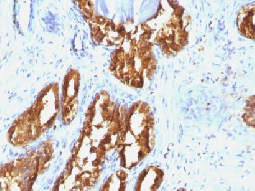
Anti-Prostate Specific Acid Phosphatase (PSAP) Antibody
Mouse Monoclonal Antibody
- SPECIFICATION
- CITATIONS
- PROTOCOLS
- BACKGROUND

Application
| IHC-P, IF, FC |
|---|---|
| Primary Accession | P15309 |
| Other Accession | 433060 |
| Reactivity | Human |
| Host | Mouse |
| Clonality | Monoclonal |
| Isotype | Mouse / IgG1, kappa |
| Clone Names | ACPP/1338 |
| Calculated MW | 44566 Da |
| Gene ID | 55 |
|---|---|
| Other Names | 5'-nucleotidase (5'-NT); Acid phosphatase prostate; ACP3; Ecto-5'-nucleotidase; Prostatic acid phosphatase (PAP); Prostatic acid phosphatase; Thiamine monophosphatase (TMPase) |
| Application Note | Flow Cytometry (0.5-1ug/million cells); Immunofluorescence (0.5-1ug/ml); ,Immunohistology (Formalin-fixed) (0.5-1.0ug/ml for 30 minutes at RT) ,(Staining of formalin-fixed tissues requires boiling tissue sections in 10mM citrate buffer, pH 6.0, for 10-20 min followed by cooling at RT for 20 minutes),Optimal dilution for a specific application should be determined. |
| Format | 200ug/ml of Ab purified from Bioreactor Concentrate by Protein A/G. Prepared in 10mM PBS with 0.05% BSA & 0.05% azide. Also available WITHOUT BSA & azide at 1.0mg/ml. |
| Storage | Store at 2 to 8°C.Antibody is stable for 24 months. |
| Precautions | Anti-Prostate Specific Acid Phosphatase (PSAP) Antibody is for research use only and not for use in diagnostic or therapeutic procedures. |
| Name | ACP3 (HGNC:125) |
|---|---|
| Synonyms | ACPP |
| Function | A non-specific tyrosine phosphatase that dephosphorylates a diverse number of substrates under acidic conditions (pH 4-6) including alkyl, aryl, and acyl orthophosphate monoesters and phosphorylated proteins (PubMed:10506173, PubMed:15280042, PubMed:20498373, PubMed:9584846). Has lipid phosphatase activity and inactivates lysophosphatidic acid in seminal plasma (PubMed:10506173, PubMed:15280042). |
| Cellular Location | [Isoform 1]: Secreted |
| Tissue Location | Highly expressed in the prostate, restricted to glandular and ductal epithelial cells. Also expressed in bladder, kidney, pancreas, lung, cervix, testis and ovary. Weak expression in a subset of pancreatic islet cells, squamous epithelia, the pilosebaceous unit, colonic neuroendocrine cells and skin adnexal structures. Low expression in prostate carcinoma cells and tissues |

Thousands of laboratories across the world have published research that depended on the performance of antibodies from Abcepta to advance their research. Check out links to articles that cite our products in major peer-reviewed journals, organized by research category.
info@abcepta.com, and receive a free "I Love Antibodies" mug.

Provided below are standard protocols that you may find useful for product applications.
Background
Recognizes a protein of 52kDa, identified as prostate specific acid phosphatase (PSAP). This enzyme catalyzes the conversion of orthophosphoric monoester to alcohol and orthophosphate. It is synthesized under androgen regulation and is secreted by the epithelial cells of the prostate gland. PSAP is found in non-neoplastic adult and fetal prostatic glands, primary and metastatic prostatic carcinomas. It shows no staining in granulocytes, osteoclasts, parietal cells of the stomach, liver cells, renal cell or breast carcinomas.
If you have used an Abcepta product and would like to share how it has performed, please click on the "Submit Review" button and provide the requested information. Our staff will examine and post your review and contact you if needed.
If you have any additional inquiries please email technical services at tech@abcepta.com.
Ordering Information
Other Products
Shipping Information
