


 Foundational characteristics of cancer include proliferation, angiogenesis, migration, evasion of apoptosis, and cellular immortality. Find key markers for these cellular processes and antibodies to detect them.
 The SUMOplot™ Analysis Program predicts and scores sumoylation sites in your protein. SUMOylation is a post-translational modification involved in various cellular processes, such as nuclear-cytosolic transport, transcriptional regulation, apoptosis, protein stability, response to stress, and progression through the cell cycle.
 The Autophagy Receptor Motif Plotter predicts and scores autophagy receptor binding sites in your protein. Identifying proteins connected to this pathway is critical to understanding the role of autophagy in physiological as well as pathological processes such as development, differentiation, neurodegenerative diseases, stress, infection, and cancer.
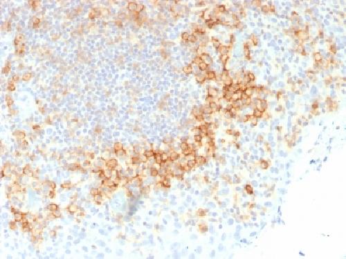
Anti-CD27 (Tumor Necrosis Factor Receptor Superfamily 7) Antibody
Mouse Monoclonal Antibody
- SPECIFICATION
- CITATIONS
- PROTOCOLS
- BACKGROUND

Application
| IHC-P, IF, FC |
|---|---|
| Primary Accession | P26842 |
| Other Accession | 355307 |
| Reactivity | Human |
| Host | Mouse |
| Clonality | Monoclonal |
| Isotype | Mouse / IgG1 |
| Clone Names | LPFS2/1611 |
| Calculated MW | 29137 Da |
| Gene ID | 939 |
|---|---|
| Other Names | LPFS2; S152; T cell activation antigen S152; T-cell activation antigen CD27; T14; TNFRSF7; TNFSF7; Tp55; Tumor necrosis factor receptor superfamily member 7 |
| Application Note | Flow Cytometry (0.5-1ug/million cells); ,Immunofluorescence (0.5-1ug/ml); ,Immunohistology (Formalin-fixed) (0.5-1ug/ml for 30 minutes at RT),(Staining of formalin-fixed tissues requires boiling tissue sections in 10mM Tris buffer with 1mM EDTA, pH 9.0, for 10-20 min followed by cooling at RT for 20 minutes),Optimal dilution for a specific application should be determined. |
| Format | 200ug/ml of Ab purified from Bioreactor Concentrate by Protein A/G. Prepared in 10mM PBS with 0.05% BSA & 0.05% azide. Also available WITHOUT BSA & azide at 1.0mg/ml. |
| Storage | Store at 2 to 8°C.Antibody is stable for 24 months. |
| Precautions | Anti-CD27 (Tumor Necrosis Factor Receptor Superfamily 7) Antibody is for research use only and not for use in diagnostic or therapeutic procedures. |
| Name | CD27 (HGNC:11922) |
|---|---|
| Function | Costimulatory immune-checkpoint receptor expressed at the surface of T-cells, NK-cells and B-cells which binds to and is activated by its ligand CD70/CD27L expressed by B-cells (PubMed:28011863). The CD70-CD27 signaling pathway mediates antigen- specific T-cell activation and expansion which in turn provides immune surveillance of B-cells (PubMed:28011863). Mechanistically, CD70 ligation activates the TRAF2-PTPN6 axis that subsequently inhibits LCK phosphorylation to promote phenotypic and transcriptional adaptations of T-cell memory (PubMed:38354704). In addition, activation by CD70 on early progenitor cells provides a negative feedback signal to leukocyte differentiation during immune activation and thus modulates hematopoiesis (By similarity). Negatively regulates the function of Th2 lymphocytes in the adipose tissue (By similarity). |
| Cellular Location | Cell membrane; Single-pass type I membrane protein |
| Tissue Location | Found in most T-lymphocytes. |

Thousands of laboratories across the world have published research that depended on the performance of antibodies from Abcepta to advance their research. Check out links to articles that cite our products in major peer-reviewed journals, organized by research category.
info@abcepta.com, and receive a free "I Love Antibodies" mug.

Provided below are standard protocols that you may find useful for product applications.
Background
Recognizes a protein of a disulfide-linked 120kDa dimer, identified as CD27. It is expressed on the majority of peripheral T cells, medullary thymocytes, memory-type B cells, and natural killer cells. It is a transmembrane phosphoglycoprotein that belongs to the tumor necrosis factor receptor (TNFR) superfamily. CD27 binds to its ligand CD70, a member of the TNF family, and induces T-cell co-stimulation and B-cell activation. It also interacts with TRAFs and is involved in activation of NFB and SAPK/JNK and induces apoptosis.
If you have used an Abcepta product and would like to share how it has performed, please click on the "Submit Review" button and provide the requested information. Our staff will examine and post your review and contact you if needed.
If you have any additional inquiries please email technical services at tech@abcepta.com.
Ordering Information
Other Products
Shipping Information
