


 Foundational characteristics of cancer include proliferation, angiogenesis, migration, evasion of apoptosis, and cellular immortality. Find key markers for these cellular processes and antibodies to detect them.
 The SUMOplot™ Analysis Program predicts and scores sumoylation sites in your protein. SUMOylation is a post-translational modification involved in various cellular processes, such as nuclear-cytosolic transport, transcriptional regulation, apoptosis, protein stability, response to stress, and progression through the cell cycle.
 The Autophagy Receptor Motif Plotter predicts and scores autophagy receptor binding sites in your protein. Identifying proteins connected to this pathway is critical to understanding the role of autophagy in physiological as well as pathological processes such as development, differentiation, neurodegenerative diseases, stress, infection, and cancer.
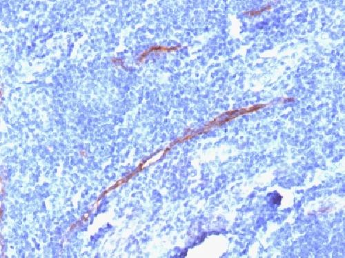
Anti-CD34 Antibody
Recombinant Rabbit Monoclonal Antibody
- SPECIFICATION
- CITATIONS
- PROTOCOLS
- BACKGROUND

Application
| WB, IHC-P, IF, FC |
|---|---|
| Primary Accession | P28906 |
| Other Accession | 374990 |
| Reactivity | Human, Rat |
| Host | Rabbit |
| Clonality | Monoclonal |
| Isotype | Rabbit / IgG, kappa |
| Clone Names | HPCA1/1806R |
| Calculated MW | 40716 Da |
| Gene ID | 947 |
|---|---|
| Other Names | Hematopoietic Progenitor Cell Antigen, HPCA1, Mucosialin |
| Application Note | Flow Cytometry (0.5-1ug/million cells); ,Immunofluorescence (0.5-1ug/ml); Western Blotting (0.5-1ug/ml);,Immunohistology (Formalin-fixed) (2-4ug/ml for 30 min at RT),(Staining of formalin-fixed tissues requires boiling tissue sections in 10mM Tris with 1mM EDTA, pH 9.0, for 10-20 min followed by cooling at RT for 20 minutes),Optimal dilution for a specific application should be determined. |
| Format | 200ug/ml of recombinant MAb purified by Protein A/G. Prepared in 10mM PBS with 0.05% BSA & 0.05% azide. Also available WITHOUT BSA & azide at 1.0mg/ml. |
| Storage | Store at 2 to 8°C.Antibody is stable for 24 months. |
| Precautions | Anti-CD34 Antibody is for research use only and not for use in diagnostic or therapeutic procedures. |
| Name | CD34 |
|---|---|
| Function | Possible adhesion molecule with a role in early hematopoiesis by mediating the attachment of stem cells to the bone marrow extracellular matrix or directly to stromal cells. Could act as a scaffold for the attachment of lineage specific glycans, allowing stem cells to bind to lectins expressed by stromal cells or other marrow components. Presents carbohydrate ligands to selectins. |
| Cellular Location | Membrane; Single-pass type I membrane protein. |
| Tissue Location | Selectively expressed on hematopoietic progenitor cells and the small vessel endothelium of a variety of tissues |

Thousands of laboratories across the world have published research that depended on the performance of antibodies from Abcepta to advance their research. Check out links to articles that cite our products in major peer-reviewed journals, organized by research category.
info@abcepta.com, and receive a free "I Love Antibodies" mug.

Provided below are standard protocols that you may find useful for product applications.
Background
This antibody recognizes a transmembrane, heavily glycosylated protein of 90-120kDa, which is identified as CD34. Its expression is a hallmark for identifying pluripotent hematopoietic stem or progenitor cells. Its expression is gradually lost as lineage committed progenitors differentiate. CD34 is a marker of choice for staining blasts in acute myeloid leukemia. In addition, it is expressed by soft tissue tumors, such as solitary fibrous tumor and gastrointestinal stromal tumor. CD34 expression is also found in vascular endothelium. Additionally, proliferating endothelial cells overexpress this molecule than the non-proliferating endothelial cells. Anti-CD34 labels > 85% of angiosarcoma and Kaposi s sarcoma, but shows low specificity.
If you have used an Abcepta product and would like to share how it has performed, please click on the "Submit Review" button and provide the requested information. Our staff will examine and post your review and contact you if needed.
If you have any additional inquiries please email technical services at tech@abcepta.com.
Ordering Information
Other Products
Shipping Information
